![]()
Prev Page--Lithology, Composition || Next Page--Utilization
Petrography of the Mineral, Croweburg, and Bevier Coals
Description of Components
The Mineral, Croweburg, and Bevier coals were studied petrographically from approximately 400 thin sections and from 22 column samples. Since the three coals show marked similarities, they are discussed here as a group rather than individually.
The outstanding characteristic of the coals is their finely banded appearance. The anthraxylon bands range from a maximum width of about 5 mm to the arbitrary lower limit of 0.017 mm and rarely exceed 1 mm. Typical bands of anthraxylon are shown in Plate 2, A and B. The relatively homogeneous nature of the anthraxylon is distinctive and aids in separating it from adjacent attrital coal. The bands are characteristically bright orange to red and in places are bounded by brilliant orange or yellow cuticular material having a serrated edge on its proximal side (Pl. 5, A and B). Anthraxylon exhibits several forms which depend on the part of the plant sectioned, the direction of the section, and the state of preservation of the material. Many bands are not continuous, but pinch out within a short distance or split into a number of finer bands due to branching or degradation. Lenticular bands are indicative of transverse sections. Some bands are double because the original stems were hollow cylinders.
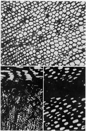
Plate 2--Thin sections showing anthraxylon bands and cell structures. A, Anthraxylon band below with attrital coal above (Bevier, X 95). B, Anthraxylon band with translucent attritus above. Attritus contains small particles of opaque pyrite and translucent calcite and clay minerals (Mineral coal X 95). C, Deformed cell structure in anthraxylon (Bevier coal X 350). D, Anthraxylon band cut parallel to bedding. Boxlike nature of cells is evident (Mineral coal (X 150). An Acrobat PDF version of this plate is available.
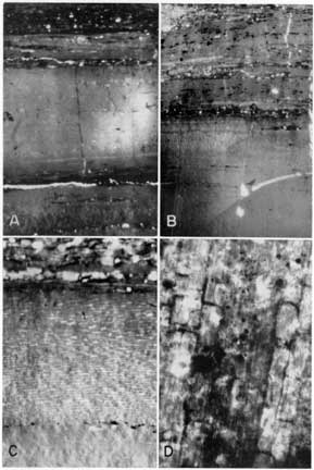
Cell structure was not seen in most sections of anthraxylon either because the plant material had undergone considerable alteration before coalification or because of lack of contrast between the cell walls and the material filling the lumens. Plate 2C is a section of anthraxylon showing traces of deformed cell structure. Plate 2D is a section of anthraxylon cut parallel to the bedding and in a direction longitudinal to the plant cells. The boxlike nature of the cells can be seen clearly.
Translucent attritus is usually heterogeneous. It consists of a closely knit debris of anthraxylon fragments, spore exines, bits of cuticle, resin bodies, and other degradation products plus extremely fine particles of calcite, pyrite, quartz, and clay minerals. Typical examples of translucent attritus are shown in Plates 3, 4, and 8C. Some translucent attritus consists entirely of small fragments which are distinguished from anthraxylon solely on the basis of size. Since most of the anthraxylon falls in the fine size range, distinction between attritus and anthraxylon is difficult. The constituents of Plate 3A are similar in appearance but the band in the middle of the section is classified as anthraxylon whereas the remainder of the material is translucent attritus.
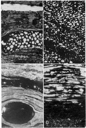
Plate 3--Thin sections showing attrital coal. A, Banded translucent attritus. Wide band near center is classified as anthraxylon on basis of size (Mineral coal, X 125). B, Translucent attritus composed of shredlike fragments. Opaque bodies are pyrite (Mineral coal, X 125). C, Opaque attritus intercalcated with bands of anthraxylon and translucent attritus (Bevier coal, X 95). An Acrobat PDF version of this plate is available.
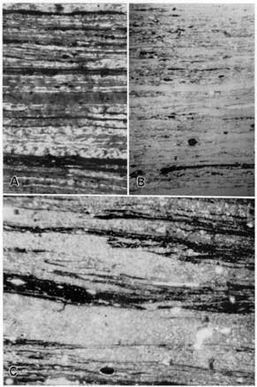
Plate 4--Resin bodies. A, Resin bodies in anthraxylon and translucent attritus. Note how bands have been compressed around resistant resin bodies (Bevier coal, X 125). B, Large resin body in translucent attritus (Bevier coal, X 125). An Acrobat PDF version of this plate is available.
Although the spore content of the coal is remarkably low, the megaspore exines can be distinguished easily when they do occur. Megaspores are brilliant yellow bodies, up to 1 mm in length, resembling a flattened tube in cross section. The ends may be invaginated as shown in Plate 5D. Plate 5C shows a cluster of megaspores at a lower magnification. The microspores are much smaller and may be mistaken for fragments of cuticle.
Bright red globular resin bodies are abundant in both translucent attritus and anthraxylon. Plate 4 illustrates how bands of anthraxylon and attritus have been compressed around more resistant resin bodies.
Plate 5--Thin sections showing cuticle and spores. A, White material is cuticle which surrounds a stem, in longitudinal section (Mineral coal, X 50). B, Fragments of cuticle. Lower double layer is a longitudinal section of a stem. Note the characteristic serrated edge on the inner side of each layer (Mineral coal, X 125). C, Cluster of megaspore exines or cases in opaque attritus (Mineral coal, X 100). D, Megaspore in opaque attritus. The double character of the spore case is clearly evident. Note the invaginated end of the case (Mineral coal, X 190). An Acrobat PDF version of this plate is available.
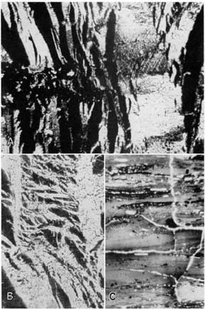
Fusain is one of the most striking components of these coals and, in places, one of the most difficult to distinguish because of its similarity to certain types of opaque attritus. At its best, it consists of cellular material with opaque cell walls and translucent spaces between the walls which have been filled with calcite. Some types of fusain are clearly transverse sections of altered wood or cortex (Pl. 6A, lower part of B, and C) since the same kinds of cells can be seen in unaltered plants. Fusain has a fibrous appearance if the cells have been cut in a longitudinal direction (Pl. 6, upper part of B) or if the original material consisted of resin rodlets. Other fusain occurs in fine irregular fragments derived from the deformation or crushing of the cellular type or from the alteration of finer woody debris. Plate 7B shows fusain in which the cell walls have been deformed so that the calcite-filled spaces no longer exhibit regularity. Fusain, in many cases, occurs in lenticular bodies (Pl. 7A).
Plate 6--Thin sections of fusain. A, Transverse section of fusinized cortex The plant cells are filled with calcite (Bevier coal, X 125). B, Upper part shows longitudinal section of fusinized plant material. Lower part shows transverse section. Cells are filled with calcite (Mineral coal, X 50). C, Transverse section of fusinized thick-walled plant cells (Bevier coal, X 125). An Acrobat PDF version of this plate is available.
Opaque attritus is a relatively minor constituent of the coals. When it does occur, it is found, in most places, near the top or bottom of the column. Clearly recognizable opaque attritus contains translucent anthraxylon-like fragments, cuticle, and spore exines in addition to the opaque constituents. Plate 3C shows opaque attritus intercalated with translucent material. Plate 5, C and D, shows examples of opaque attritus containing spore exines and fragments of cuticular material.
The most perplexing problem in the petrographic analysis of the coal was the differentiation of the opaque or semiopaque constituents. Opaque attritus may have a transitional relation with translucent attritus or anthraxylon. In such cases, the translucent constituents become progressively more opaque and grade into fusainlike material. Plate 7D is a typical example of this transition. The material at the bottom of the section is clearly anthraxylon. It changes upward into semiopaque matter which in turn becomes progressively more opaque and acquires the expanded structure of fusain. Since the boundaries of these constituents are not denned clearly, the analytical results are dependent, to a large measure, on the judgment of the observer. Plate 7C shows that other coal constituents may alter to fusain. The round opaque body is a fusinized resin globule.
Plate 7--Thin sections of fusain. A, Section of fusain lens in translucent attritus (Mineral coal, X 100). B, Fusain showing deformed plant cells (Mineral coal, X 125). C, Opaque body in lower part is a fusuinized resin globule; opaque band above is fusain (Mineral coal, X 135). D, Material at bottom is anthraxylon. Note the change upward into opaque attritus and fusuain (Mineral coal, X 50). An Acrobat PDF version of this plate is available.
Origin of Opaque Components
Because the opaque components are of considerable importance in both coal classification and coal utilization, it is pertinent to review briefly the ideas concerning the origin of these constituents. In general, two schools of thought have existed (Hendricks, 1945, pp. 19-21) concerning fusain. The first attributed the origin to forest fires, and the second to some form of chemical alteration prior to burial. The forest-fire theory has found little favor because of the improbability of extensive fires in typical peat swamps and the absence of ash layers. In addition, fragile plant structures are fusinized and plant stems are found with fusain on the interior. The chemical alteration theories include dehydration during periods of dryness or by sulfuric acid, carbonization as the result of catalytic action, impregnation with various salts or gases which promote carbonization and inhibit aerobic decay, and the local action of thermophilic bacteria.
The origin of opaque attritus has received relatively little attention and most investigators have considered it a separate entity from fusain. Thiessen and Sprunk (1936) observed a transitional relation between opaque and translucent constituents in their studies of the Upper and Lower Cedar Grove coals of West Virginia. Fieldner and Schmidt (1941, p. 12) suggested that opaque attritus resulted from advanced decomposition due to the action of biological agencies and that as decay progressed, the attritus became more opaque.
Certain observations may be made concerning the origin of the opaque constituents as illustrated in Kansas coals.
(1) The process or processes which produce fusain may act upon any of the plant materials and the resulting degree of opacity is dependent upon the intensity and duration of the process. In addition, it is likely that certain plant tissues are more susceptible to fusinization than others. Thin sections show the gradation of anthraxylon and translucent attritus into opaque attritus and fusain. It seems probable that had the process producing the gradation been of sufficient intensity and duration, all the material would have been fusinized. Since fusinized resin bodies have been found, it is evident that even the more resistant plant materials are susceptible to fusinization.
(2) The process of fusinization was operative in the early stages of coalification. Cellular fusain is able to maintain its open structure because the cells are filled with calcite. The other coal material is seen to be compressed around the structurally supported fusain. Therefore fusinization and impregnation took place prior to compaction of the surrounding coal and the process may be regarded as diagenetic. If coalification had proceeded in a normal manner, cell cavities would have filled with humic material and later fusinization would have produced a homogeneous rather than a cellular fusain.
(3) Fusinization can be local in nature. Most fusain in Kansas coals cannot be traced for any distance laterally whereas, in some coals, fusain bands are persistent. It would seem that fusain is produced by a number of processes, some of which are local in action whereas others are not.
(4) Much that has been classed as opaque attritus is probably crushed and compacted fusain. In cases where no calcite impregnation of cells took place, the structurally weak fusain was not able to maintain its open spaces. Undoubtedly much of the crushed fusain has been classified as opaque attritus since fusain is identified largely on the basis of its open cell structure.
Analysis of Components
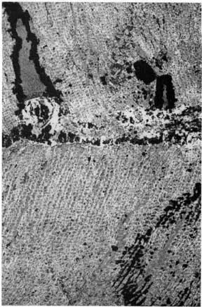
In Plate 1, the type of coal and the percentage of components in each thin section have been shown graphically for 22 column samples. As illustrated in Plate 1A, the type of coal is designated at the left side of the figure. Distances from the top of the bed are given at the left margin. The thickness of each lithologic unit and its description number are shown in the next two columns. The percentage loss in the lower left corner is an indication of the amount of coal lost through sawing and grinding. It was determined by dividing the thickness of the column sample after grinding (as determined from the sum of the thin section widths) by the original thickness of the column before grinding. Approximately true thickness can be found by multiplying the diagram thickness by the percent loss and adding the product to the diagram thickness. The percentages of anthraxylon, translucent attritus, opaque attritus, and fusain are shown by bar diagram to the right of the figure.
Mineral Coal
Petrographic analyses of the Mineral coal are shown in Plate 1, A to G. With few exceptions, the Mineral coal is a uniformly bright coal. It is characterized by a relatively large content of translucent attritus. Opaque attritus and fusain are minor constituents. In the few cases where opaque attritus increases to the extent that parts of the coal are classified as semisplint or cannel, these constituents are confined to the top or bottom of the bed. Sample Ck-3-M (Pl. 1C) contains 0.8 inch of semisplint at the top of the column. Sample Cr-8-M (Pl. 1D) has 0.6 inch of semisplint about 1.4 inches from the top and another semisplint band 0.8 inch wide at the bottom of the bed. Sample Ck-5-M (Pl. 1E) is somewhat peculiar in that it contains a thin band of cannel coal at the bottom of the bed. Although this band meets the petrographic requirements of a cannel, it does not have the typical compact and nonbanded canneloid appearance. About 1.4 inches of the column is missing since part of the coal was too friable to section or polish.
Banded or nodular pyrite is relatively rare in the Mineral coal except at the south end of the field. Although these impurities are described as pyrite in the diagrams, they are actually "coal balls" and contain considerable calcite. Samples Ck-4-M (Pl. 1F) and Ck-2-M (Pl. 1G) both show that the nodules are confined to the upper portion of the coal bed.
The average analysis for each column is tabulated in Table 9 and the average of these analyses is shown at the bottom of the table. It is interesting to note that opaque attritus increases from north to south until it reaches a maximum of 9.0 percent just north of Frontenac (sample Cr-8-M) and then decreases to the south. This variation may not be significant since relatively few Mineral coal samples have been analyzed. Nevertheless, the opaque attritus content of the coal is lower in the north and south ends of the field.
Table 9--Summary of data on petrographic analyses of column samples of coals from the Southeastern Kansas coal field
| Locality no. |
Sample no. |
Thickness, inches |
Anthraxylon, percent |
Translucent attritus, percent |
Opaque attritus, percent |
Fusain, percent |
|---|---|---|---|---|---|---|
| Mineral coal | ||||||
| 1 | Cr-2-M | 10.4 | 26.1 | 66.9 | 2.6 | 4.4 |
| 2 | Cr-4-M | 13.5 | 21.3 | 69.0 | 1.4 | 8.2 |
| 3 | Cr-3-M | 18.5 | 24.1 | 64.3 | 3.9 | 7.6 |
| 9 | Cr-8-M | 21.1 | 33.5 | 50.9 | 9.0 | 10.3 |
| 15 | Ck-5-M | 14.3 | 31.2 | 59.4 | 2.7 | 6.6 |
| 16 | Ck-4-M | 18.4 | 41.1 | 51.6 | 1.4 | 5.9 |
| 18 | Ck-2-M | 10.4 | 34.4 | 58.6 | 1.5 | 5.5 |
| Average | 15.2 | 30.2 | 60.1 | 3.2 | 6.9 | |
| Bevier coal | ||||||
| 19 | Bn-3-B | 12.9 | 45.5 | 40.4 | 8.0 | 6.1 |
| 20 | Bn-2-B | 14.0 | 54.4 | 35.4 | 4.8 | 5.4 |
| 21 | Bn-1-B | 12.1 | 41.7 | 40.7 | 1.6 | 16.0 |
| 22 | Cr-9-B | 15.9 | 38.3 | 53.8 | 4.4 | 3.5 |
| 25 | Cr-5-B | 16.8 | 27.5 | 53.9 | 4.2 | 14.4 |
| 26 | Cr-6-B | 17.5 | 33.7 | 55.5 | 5.8 | 5.1 |
| 27 | Cr-7-B | 15.0 | 36.2 | 55.6 | 4.5 | 3.8 |
| 30 | Cr-12-B | 16.1 | 36.1 | 52.7 | 4.4 | 6.8 |
| 31 | Cr-14-B | 15.4 | 32.0 | 53.7 | 9.7 | 4.7 |
| 32 | Cr-11-B | 14.1 | 36.8 | 53.1 | 5.1 | 5.0 |
| 33 | Cr-13-B | 15.4 | 35.4 | 49.8 | 4.2 | 10.5 |
| 34 | Cr-10-B | 15.7 | 31.7 | 55.7 | 6.9 | 5.7 |
| 35 | CK-1-B | 13.4 | 36.2 | 50.3 | 8.2 | 53 |
| 37 | Lt-1-B | 12.0 | 44.4 | 38.2 | 11.4 | 6.0 |
| Average | 14.7 | 37.9 | 49-2 | 5.9 | 7.0 | |
| Croweburg coal | ||||||
| 8 | Cr-1-C | 11.5 | 21.0 | 58.2 | 7.7 | 13.1 |
Bevier Coal
Petrographic analyses of the Bevier coal are shown in Plate 1, H to U. The Bevier is also a uniformly bright coal except for a few thin bands of splint and semisplint coal. Translucent attritus again predominates whereas opaque attritus and fusain are relatively minor constituuents. No splint or semisplint coal is encountered north of Franklin in Crawford County. The first occurrence is in sample Cr-5-B (Pl. 1L) which has a thin band of semisplint near the top of the bed. There is also a layer of almost pure fusain about 11.5 inches from the top of the bed which does not fit into the type classification scheme. Other samples to the south show small amounts of splint or semisplint near the top or bottom of the bed. Samples Cr-14-B (Pl. 1P) and Cr-ll-B (Pl. 1Q) are exceptions to this general rule since semisplint coal occurs near the middle of the bed.
Nodular or banded pyrite is rare in the Bevier coal. The only occurrence was in sample Ck-1-B (Pl. 1T) about 8 inches from the top of the sample.
The average analysis for each column sample is tabulated in Table 9 and the average of the analyses for all the columns is shown at the bottom of the table. The distribution of components is somewhat erratic and there is no apparent systematic variation.
Croweburg Coal
A single analysis of the Croweburg coal is shown in Plate 1V. It is similar in most respects to the Mineral and Bevier coals. The most abundant constituent is translucent attritus. A relatively thick band of splint coal occurs near the bottom of the bed. The average analysis of the column is tabulated in the lower part of Table 9.
Comparison of the Coals
In comparing the analyses of the coals, it is important to note that the Bevier coal is somewhat higher in anthraxylon, opaque attritus, and fusain than the Mineral coal. The economic significance of these differences will be discussed later in reference to coal utilization. Since the distribution of splint and semisplint coal is similar for all the beds, it may be pertinent to mention that one of the advantages of column analysis is to show the distribution of coal types so that selective mining may be employed intelligently. The value of such a practice in mining Kansas coals is doubtful, however, because of the thinness of the beds and the relatively small contribution of the splints and semisplints to the overall character of the coal.
Mineral Matter
Chemical analyses of the Mineral, Croweburg, and Bevier coals already have indicated their relatively high ash content. However, the ash value contributes little to an understanding of the identity and distribution of the mineral matter which is responsible for the ash. Such information is important for several reasons: (1) it aids in determining the extent of economical coal beneficiation; (2) the chemical and physical properties of coal are influenced by the character of the mineral matter; (3) it is a reflection of the geologic history of the coal bed.
Kinds of Mineral Matter
Two kinds of mineral matter are generally found in all coals. The first is called "inherent mineral matter" and refers to that portion organically combined with the coal. It cannot be determined petrographically and includes those elements which have been assimilated by plants for nutritive purposes. The second is known as "extraneous mineral matter" and includes that portion which is foreign to the plant material. Extraneous mineral matter is the larger contributor to the total ash content. It consists of detrital minerals deposited during coal accumulation and minerals deposited from solution or suspension during and after coal accumulation. Most of the mineral matter in Kansas coal is of the latter type.
Method of Study
The techniques used in the study of the mineral constituents are outlined as follows:
(1) Approximately a 25 gram sample, crushed to minus 60 mesh size, was split from each of the channel samples used for chemical analysis. After weighing, the sample was placed in a large separatory funnel containing a 1.70 specific gravity solution of bromoform and carbon tetrachloride. Upon complete separation, the float-sink fractions were collected on filter paper, washed with acetone, dried, and weighed.
(2) The sink fraction was placed in a 2.90 specific gravity solution of tetrabrom-ethane and after separation, the float-sink fractions were collected and washed.
(3) Part of the 2.90 sink fraction was mixed with bakelite powder and mounted in a bakelite disc for polishing. The polished section was studied with a reflecting microscope. Color, hardness, internal reflection, and anisotropism were used to identify the opaque minerals.
(4) Both the float and sink fractions from the 2.90 specific gravity separation were examined with the aid of binocular, petrographic, and reflecting microscopes. Hydrochloric acid solubility, flame reactions, optical properties, and microchemical tests were used to identify the minerals.
(5) The thin sections prepared for column analysis were used to study the form and distribution of the minerals.
(6) Polished sections of pyrite nodules were made for study with the reflecting microscope and peels of the "coal balls" were made.
Mineral Assemblage and Distribution
Although the mineral content of Kansas coals is high, the kinds of minerals are few. Calcite and pyrite are the most important constituents. There are also lesser amounts of aragonite, marcasite, sphalerite, quartz, apatite, and clay minerals.
Detrital minerals--The only known detrital minerals in the coals are quartz, apatite, and clay minerals. They are found mainly in the attrital portions of the coal and along bedding planes. A number of well-rounded grains of quartz and apatite were seen in the float-sink fractions of several samples. Clay minerals are the most abundant detrital constituents.
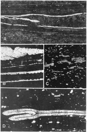
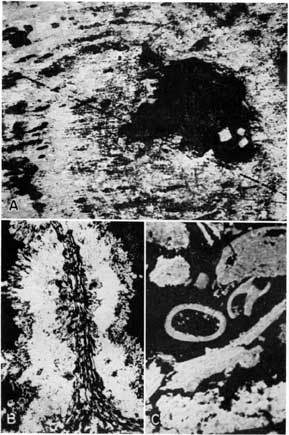
Calcite--Calcite is the most important mineral constituent of the coals. It has two different modes of occurrence. The largest amount of calcite is found in cleats and dessication fractures where it may be associated with aragonite. The fractures usually traverse the coal normal to the bedding and may exhibit intricate, branching patterns as shown in Plate 8 A and B. Aragonite was identified in the crushed samples by its orange color, fibrous appearance, and refractive index. In thin sections, the aragonite resembles calcite but usually can be distinguished by its biaxial interference figure and fibrous structure. It may grade into coarsely crystalline calcite which shows undulatory extinction, indicating that at least some of the calcite has resulted from an aragonite-calcite transition. Little is known of the stability range or the conditions favoring the precipitation of aragonite. Rankama and Sahama (1950, p. 470) state that ground water often precipitates calcium, carbonate either as calcite or aragonite. The stability of aragonite is probably a sensitive function of the partial pressure of carbon dioxide, temperature, and pH. The fracture-filling type of carbonate probably was deposited after coalification since it is unlikely that such fractures could develop in plastic peat.
Plate 8--Thin sections of mineral matter in coal. A, Intricate pattern of calcite in minute fractures in coal, under crossed nicols. Note cleavage and twinning in calcite (Mineral coal, X 125). B, Calcite filling fractures in coal (Mineral coal, X 125). C, Section shows distribution of minerals along banding in translucent attritus. Small opaque bodies are pyrite; lighter bodies consist largeley of clay minerals Notice the cube of pyrite in the upper right corner (Mineral coal, X 120). An Acrobat PDF version of this plate is available.
Calcite also occurs in the open spaces of fusain (Pls. 6 and 7) and as the impregnating material of "coal balls." This calcite probably was deposited prior to coalification since it provides structural support for the cells which could not have maintained their form unless they were filled before compaction of the surrounding coal. This type of calcite is easily identified in both the crushed samples and the thin sections. In the crushed samples, the cell walls are often broken away from the calcite so that it emerges as a cast of the cell interior and looks like a small, milled cylinder.
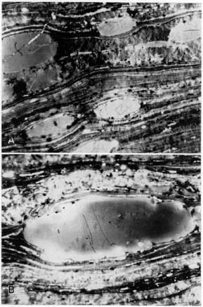
The "coal balls," which are found only in the Mineral coal, are of particular interest. They are nodules or laterally persistent bands of calcite-impregnated plant material. Since the original woody parts of the plant are preserved in the calcite, they have been an important source of information concerning the structure of coal-forming plants. Plate 9 shows two photomicrographs of polished "coal balls" taken with the aid of a reflecting microscope using obliquely incident light. Plate 10 is a projection print of a peel of the same material. In both cases, the original cell walls can be seen clearly.
Plate 9--Calcite in "coal balls." A, Photomicrograph of a coal ball taken with a reflecting microscope using obliquely incident light. The material in the center of the boxlike cells is calcite; the original woody plant material is preserved in cell walls (X 80). B, Same, showing different section of plant cells (X 85). An Acrobat PDF version of this plate is available.
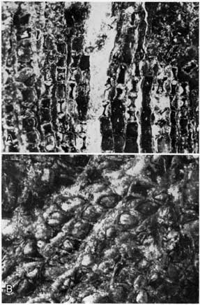
Plate 10--"Coal ball" peel. The woody plant material is preserved in calcite and can be seen distinctly. Black material is coal (Mineral coal, X 16). An Acrobat PDF version of this plate is available.
Pyrite--Pyrite is widely distributed throughout the coal beds and is occasionally associated with marcasite. It occurs principally as disseminations in the attrital parts of the coal as shown in Plate 8C. Well-developed cubes like the one in the upper right corner of the photograph can be seen in some thin sections. The small opaque particles in the photograph are also pyrite.
Although quantitatively less important, the most interesting occurrence of pyrite is in nodules and bands. The distribution of these forms was shown earlier in the profile diagrams of the column samples. The nodules and bands of the Kansas coals are never simple, concretionary forms deposited along bedding planes, but are intimately associated with the plant material. In most cases, the pyrite replaces calcite which had either filled fusain open spaces or impregnated "coal balls." In other cases, the pyrite completely replaces the entire plant or coal material so that evidences of earlier calcite are entirely lacking. In some places, transitions from partially replaced calcite and plant material to massive pyrite are found. Relations of this kind were observed in polished sections. Plate 11 shows pyrite replacing the calcite filling of plant cells. In Plate 11A and B, the cell structure is preserved intact, for the most part, so that each cell is individually outlined in pyrite. Toward the lower left corner of Plate 11A, there are areas of more complete replacement. The white material in the photographs is pyrite, the dark partitions between the blebs of pyrite are preserved cell walls, and the remainder of the dark material is principally calcite. Plate 11C shows replacement of both calcite and coal. The areas of pyrite have begun to coalesce where calcite and coal are replaced. Plate 11D shows partial replacement and also illustrates minor deformation of the plant structure. Pyritic replacement of individual plant parts is illustrated in Plate 12. A transverse section of a stem in Plate 12A shows almost complete replacement of the peripheral part and only partial replacement of the central portion. Plate 12B is a longitudinal section of a stem which again shows only partial replacement in the pithy part of the plant. The difference in the character of the pith cells and the wood and cortex cells is rather well illustrated. Plate 12C shows a mixture of replaced plant debris. In one place, a transverse section of a replaced stem can be seen.
Plate 11--Pyrite in coal. Photomicrographs of polished sections taken with the reflecting microscope using vertically incident light. A, White material is pyrite; black material is calcite and coal. Pyrite has replaced calcite which originally filled cell cavities and has partially replaced coal (Mineral coal, X 95). B, Pyritic replacement of calcite at higher magnification (Mineral coal, X 375). C, Coalescence of pyrite where it replaces calcite and coal (Mineral coal, X 95). D, Pyritic replacement of calcite and coal. Plant cells have been somewhat deformed (Mineral coal, X 95). An Acrobat PDF version of this plate is available.
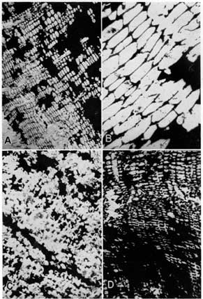
Plate 12--Pyrite in coal. Photomicrographs of polished sections taken with the reflecting microscope using vertically incident light. A, Transverse section of a plant stem replaced by pyrite. White material is pyrite. The stem is almost completely replaced at its periphery but is only partially repalced at the center (Mineral coal, X 95). B, Longitudinal section showing same relation as above (Mineral coal, X 95). C, Pyritized plant debris. Note transverse section in center (Mineral coal, X 95). An Acrobat PDF version of this plate is available.
The origin of pyrite is somewhat problematic. However, most authors agree (see Rankama and Sahama, 1950, p. 668) that iron sulfide is precipitated in stagnant water in the presence of hydrogen sulfide. The processes which produce hydrogen sulfide are bacterial reduction of sulfoproteins, bacterial reduction of sulfates, and bacterial action on free sulfur. Dissolved iron compounds such as ferrous sulfate, as well as colloidal and precipitated ferric hydroxide, react with the hydrogen sulfide to form iron sulfide. Marcasite was identified in a few places. The presence of marcasite is not too surprising in the light of Buerger's (1934) studies of the pyrite-marcasite relation. Buerger points out that pyrite and marcasite can precipitate together--the proportion of marcasite being a function of the hydrogen ion concentration (e.g., the lower the pH, the greater the amount of marcasite).
Sphalerite--Sphalerite is a minor constituent of the coals. Traces of it were found in the sink fractions of all but a few of the samples. No sphalerite was positively identified in the thin sections. This could easily be due to the loss of such a constituent in cutting to a thickness of 10 microns since much of the calcite could not even be saved. The sphalerite distribution can only be inferred from the angularity of the fragments in the crushed samples. In all probability it was deposited in the calcite-filled cleats and fractures.
The distribution of the mineral matter in the coal has historical significance in that it has served to indicate something of the origin of fusain. It might also be inferred from the extremely low content of detrital minerals in Kansas coals that extremely stable conditions prevailed during the time of coal accumulation.
Prev Page--Lithology, Composition || Next Page--Utilization
Kansas Geological Survey, Geology
Placed on web November 2005; originally published May 1953.
Comments to webadmin@kgs.ku.edu
The URL for this page is http://www.kgs.ku.edu/Publications/Bulletins/102_1/06_petro.html