![]()
Prev Page--Biostratigraphy || Next Page--Paleoecology
Petrology
Shaly Chalk
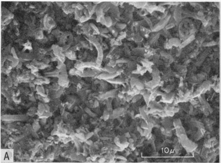
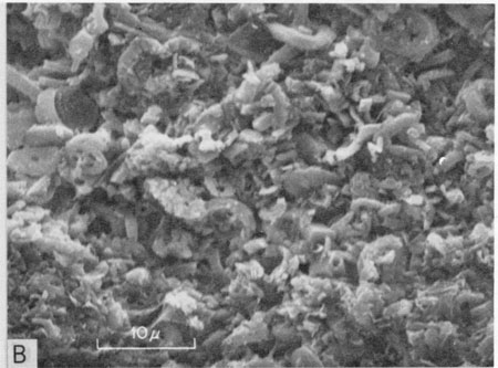
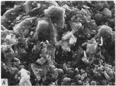
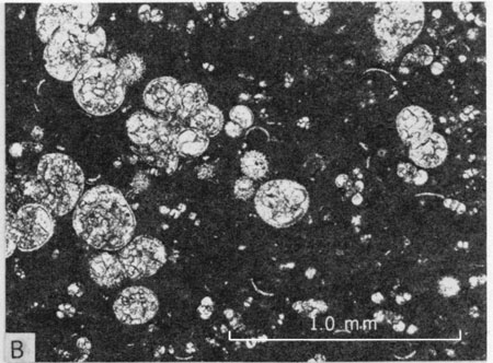
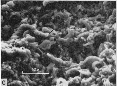
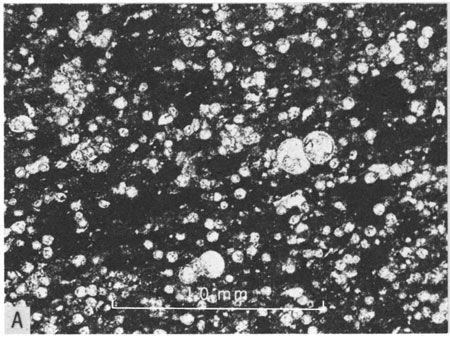
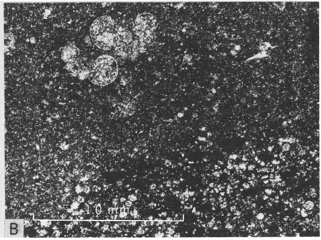
Throughout the Greenhorn Limestone of Kansas the most common lithology is shaly chalk; samples of this from throughout the section were studied in thin section. Shaly chalk is a micritic rock consisting of poorly lithified calcitic mud together with wide ranging amounts of allochems, organic matter, micrograined terrigenous detritus, and secondary minerals such as pyrite, hematite or limonite. The matrix is composed of grains less than 5µ in size and has a minutely grainy texture. Under plane-polarized light the color of the matrix is usually grayish orange to pale brown or yellowish gray but is darker shades of orange and brown where stained by organic matter or iron oxide. Scanning electron microscopy reveals that coccoliths are the principal component of the shaly chalk matrix (Fig. 16,A,B). Hattin and Darko (1971, p. 8) reported that some Greenhorn shaly chalk samples contain as many as 6 X 10⁹ coccoliths per cm3. Shaly chalk includes both packstone and wackestone depositional textures.
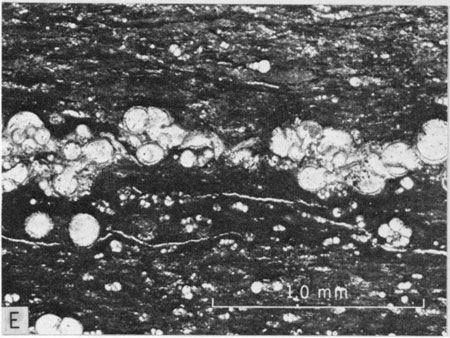
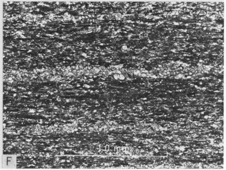
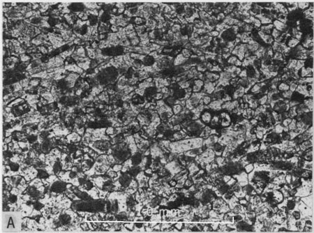
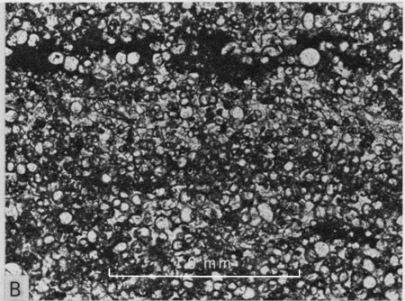
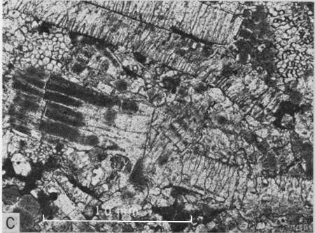
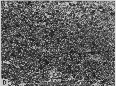
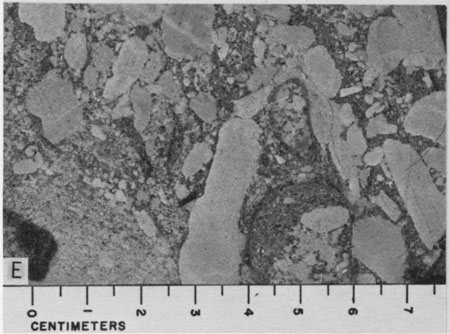
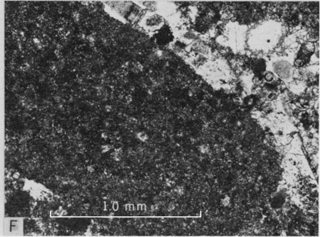
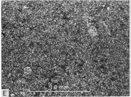
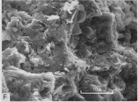
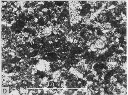
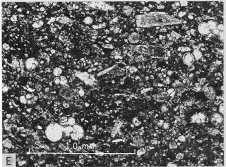
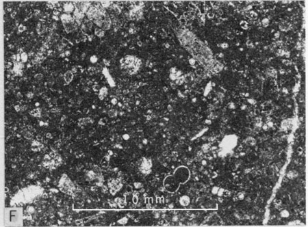
Figure 16--Petrologic features of the shaly chalk. A) Scanning electron micrograph of shaly chalk from middle part of Lincoln Member at Locality 1 showing poorly cemented character of coccolith-rich shaly chalk [bar is 10 µ]. B) Same, lower part of Hartland Member at Locality 13 [bar is 10 µ]. C) Photomicrograph of shaly chalk thin section from upper part of Lincoln Member at Locality 3 showing abundance of horizontally elongated, lenticular fecal pellets (gray bodies). Light-colored laminae are composed mostly of foraminifer tests. Plane-polarized light [bar is 10 mm]. D) Photomicrograph of shaly chalk thin section from middle part of Lincoln Member at Locality 6 showing lamina composed mostly of compacted fecal pellets in lower half of photo. Plane-polarized light [bar is 10 mm]. E) Photomicrograph of shaly chalk thin section from upper half of Hartland equivalent of Bridge Creek Member at Locality 14 showing lamina composed of large foraminifer tests. Note coarse spar calcite in chambers of tests. Plane-polarized light [bar is 10 mm]. F) Photomicrograph of shaly chalk from lower part of Pfeifer equivalent of Bridge Creek Member at Locality 14 showing two very thin laminae composed predominantly of silt-size Inoceramus prisms. Note abundant wispy grains of organic matter (black) oriented parallel to bedding. Plane-polarized light [bar is 10 mm].
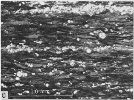
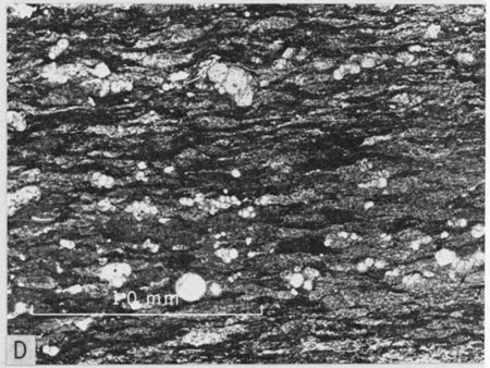
In addition to coccoliths the shaly chalk is characterized also by a variety of allochems. Stratigraphic variation in concentration of allochems accounts for much of the lamination that is evident in most exposures of shaly chalk. One of the two dominant allochems is fecal pellets (Fig. 16, C). These have been flattened during compaction and are fusiform in vertical section. Together with other elongate particles like fish scales and bits of organic matter the pellets commonly arch over or bend beneath more resistant grains including foraminifer tests and Inoceramus fragments. In plane-polarized light the pellets are light gray to yellowish gray, are nearly white in reflected light, and have a grainy texture. Pellets can be distinguished readily from the matrix by shape, lighter and more uniform color, and more homogeneous. texture. Pellet borders are usually defined sharply so that pellets are in marked contrast to the adjacent matrix. However, many pellets have indistinct borders so that contact with the adjacent matrix is not easily detected. Pellets range greatly in size; the smallest are in the coarse silt range. The largest are as much as 0.5 to 0.6 mm in long dimension, but such wide specimens have resulted from lateral spreading during compaction and are very thin in proportion to length. Less-compacted pellets, having the outline of a football, are as much as 0.1 mm in thickness in specimens only 0.25 mm in long dimension. Because of great abundance these flattened pellets contribute significantly to the fissility of shaly chalk beds. Concentrations of fecal pellets are a common cause of lamination in shaly chalk (Fig. 16,D).
Equally important allochems include foraminifer tests; these are ubiquitous constituents of shaly chalk. In a recent study of the Greenhorn Limestone, Eicher and Worstell (1970) reported 22 species of planktonic foraminifera from shaly chalk beds at Locality 3 and 20 species from Localities 13 and 14. The most abundant species are Heterohelix globulosa (Ehrenberg), Hedbergella delrioensis (Carsey), H. planispira (Tappan), H. amabilis Loeblich and Tappan, H. portsdownensis (Williams-Mitchell), and Whiteinella aprica (Loeblich and Tappan). They found benthonic foraminifera to be generally rare in the formation.
Greenhorn foraminifera display a wide range in quality of preservation. At one extreme are tests that have been preserved intact, including details of pores and spines; at the other are specimens in which the chamber walls have been partially or wholly obliterated by diagenetic processes. Many tests are broken, some by compaction, others by preburial processes as evident from isolated test fragments. Partial solution of tests has occurred along some grain-to-grain contacts, and some tests are truncated by solution contact with the adjacent matrix. The tests are composed of calcite. Size of tests ranges widely, even within a single thin section. The smallest specimens are in the coarse silt range, the largest are at the small end of the coarse sand range. A majority of all multichambered specimens fall in the very fine sand to medium sand range. The chambers of these tests are filled with sparry calcite occurring usually as a single crystal that joins the test wall without indication that the chambers were filled by inward growth of numerous fringing crystals. In some specimens, the spar in all chambers is in optical continuity and apparently represents a single large crystal. In a small number of specimens, one or more chambers are filled by more than one calcite crystal, but the number rarely exceeds four. Cleavage planes are visible in much of the chamber-filling calcite. Locally silica has replaced calcite in the chambers of some foraminifera.
In some thin sections of shaly chalk foraminifer tests are sparse; in others the rock is crowded with tests scattered irregularly through the rock. Tests concentrated in discrete layers are a common cause of even lamination in shaly chalk (Figs. 16,C,E). These laminae range from layers without stacking of tests and as little as 0.06 mm in thickness to laminae having tests stacked several high and 0.5 mm or more in thickness. In general, the thinnest laminae are composed of silt-size foraminifera whereas thicker laminae are composed of larger tests. The laminae are cemented by patches of sparry calcite but patches of micrite are commonly incorporated. Thin, even laminae of these types are more abundant in the Lincoln Member and upper part of the Hartland Member. Some foraminifer-rich laminae also contain pellets and other debris, all set in a micritic matrix. These represent less well-washed concentrations of tests. In addition to the thin, even laminae described above, some shaly chalk thin sections contain thicker, irregular laminae of foraminifer tests that also include Inoceramus prisms and valve fragments. These laminae are several millimeters in thickness and are actually thin lenses of biosparite which are described in detail in the next section. All of the well-washed concentrations of foraminifer tests are the result of very gentle current action that winnowed bottom sediments at irregular intervals.
Isolated prisms, valves, and valve fragments of Inoceramus are common in shaly chalk beds throughout the Greenhorn. The aragonitic nacre of such valves is not present in any thin sections studied by me. Valves or valve fragments rarely exceed 1 mm in maximum thickness; prisms range in diameter from silt to fine sand size. Tabular valve fragments and isolated prisms have markedly preferred orientation more or less parallel to general stratification; most such specimens consist of unrecrystallized calcite so that the prismatic structure is preserved in fine detail and growth lamellae are commonly visible also. In some thin sections irregular masses of pyrite have replaced small areas of some Inoceramus valve fragments. In a majority of specimens the Inoceramus grains are in sharp contrast with the adjacent matrix; a few grains have notched borders that have been infilled by the matrix. Partial solution has occurred along some grain-to-grain contacts, whether between Inoceramus grains or between Inoceramus and foraminifer tests. Some of these solution contacts are notched or sutured.
Inoceramus grains are common in laminae composed dominantly of foraminiferal tests; sparse, very thin laminae consist predominantly of Inoceramus grains (Fig. 16,F). Such laminae contain few to many foraminifer tests and are cemented by sparry calcite. In most shaly chalk samples Inoceramus grains are less abundant than either foraminifer tests or pellets.
Fragments of oyster valves were recorded in a few thin sections but these are rare. In one such sample a single valve fragment is partially silicified.
Shaly chalk samples also contain from less than 1 percent to approximately 10 percent matter having questionable composition. Minute, lenticular to wispy bodies of opaque material are scattered throughout most thin sections and are aligned parallel to general stratification (Fig. 16,D,E,F). Although some of these bodies are as much as 0.8 mm in length the vast majority are less than 0.25 mm long. The opaque bodies have a thickness maximum of about 0.1 mm but some of the longest bodies are less than half that thickness. These bodies are commonly surrounded by matrix that has been stained dark reddish brown, apparently by substances derived from this problematic material. The matrix is more heavily stained in shaly chalks having greatest abundance of these bodies. Like the pellets described above, these bodies apparently have suffered much compaction and are commonly draped over, or bent downward beneath, resistant skeletal grains. In some rocks concentrations of these bodies help to form yet another type of lamination, but laminae of this kind are not common. These bodies may be of multiple origin. The most obvious possibilities include a) inorganic matter consisting in part of terrigenous detritus, b) carbonaceous matter, and c) organic matter other than carbonaceous material. Although the many shaly chalk samples subjected to insoluble residue analysis contain from 9.5 to 56.6 percent insoluble residue, and although these residues consist largely of microscopic grains of terrigenous detritus (Hattin, 1971, p. 423), the bodies described here have shape and size that is difficult to reconcile with ordinary deposition of terrigenous detritus. The latter should occur as thin layers or as grains scattered throughout the rock, not as discrete lenticular bodies. Furthermore, the black bodies are opaque; silt and sand-size grains of terrigenous detritus would mostly be non-opaque. Chemical analyses of little-weathered shaly chalk samples contain 0.37 to 6.3 percent organic carbon, averaging 3.3 percent for 20 analyses. Only three of these values are below 2.3 percent. The range in percentage of opaque bodies in thin sections is compatible with the percentage of organic matter determined by chemical analysis. I have concluded that the opaque bodies are composed of some kind of organic matter. The reddish discoloration of matrix associated with the opaque bodies suggest iron oxide staining and high iron content in the bodies. If these bodies are organic, as seems likely, they may contain iron as a by-product of organic decay.
Skeletal remains of fish, and possibly other vertebrates, are evident in all thin sections studied. These bone fragments and scales are bright yellow- to deep amber-colored in plane-polarized light and are gray or opaque under crossed nicols. Most grains are elongate and oriented parallel to bedding. Such grains range widely in size and are scattered through the matrix and also occur sparingly in foraminiferal laminae.
The HCl-insoluble fraction of shaly chalks mostly is not obvious in thin section. The residues examined consist mainly of micrograined terrigenous detritus that apparently is disseminated throughout the rock matrix. Iron compounds, including pyrite, hematite, and limonite are second in abundance; these occur as partial replacement of skeletal debris, as silt-size blebs inside chambers of foraminifer tests in some thin sections, scattered through the matrix, and apparently also as a component of the opaque organic bodies described above. Chemical analysis of 19 samples of nonweathered, nonbentonitic shaly chalk gave a range in pyrite content from 0.64 to 6.94 percent, averaging 2.0 percent. In all but one pair of samples analyzed, pyrite content of shaly chalk is greater than that of the adjacent bed of chalky limestone. A few residues contain partially silicified Inoceramus fragments and/or siliceous replacements of the calcite filling of foraminifer chambers. Organic matter, sparse gypsum crystals, and silica make up the remainder of these residues. Percentage of residue in shaly chalk samples averages 31.9 percent for 88 samples. In a detailed study of Hartland, Jetmore, and Bridge Creek strata the writer (Hattin, 1971, p. 425, 426) determined that residue percentages generally decrease upward in the Greenhorn section and increase generally in a westward direction.
Biosparite
Lincoln Member
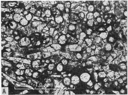
Skeletal limestones of the Lincoln Member were studied under the petrographic microscope and classified into three major categories. These are, in order of decreasing abundance, Inoceramus-prism biosparite (Fig. 17,A) foraminiferal biosparite, (Fig. 17,B) and Inoceramus-fragment biosparrudite (Fig. 17,C). The principal constituents of each occur also in the other kinds of rock. In all three types of rock the Inoceramus prisms occur as single-crystal prisms derived from disintegration of the prismatic layers of Inoceramus valves. Prism diameters range from coarse silt to medium sand and reach lengths as great as two mm. In a majority of thin sections prisms have a strong preferred orientation parallel to stratification or cross-stratification planes. The prisms are generally in sharp, even contact with interstitial cement, but all thin sections contain numerous prisms exhibiting peripheral alteration that is related to the cementation process; parts of a given prism appear to have been invaded by calcite cement. A smaller number of prisms have an indistinct or blurred border where in contact with cement, or more commonly where in contact with micritic matrix that occurs locally in these predominantly sparry-calcite-cemented rocks. A few prisms exhibit sutured or notched contacts where in contact with adjacent prisms.
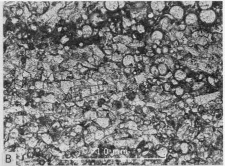
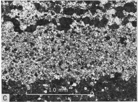
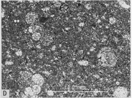
Figure 17--Petrologic features of skeletal and intraclastic limestone. A) Photomicrograph of Inoceramus-prism biosparite from middle part of Lincoln Member at Locality 1. Plane-polarized light [bar is 1.0 mm]. B) Photomicrograph of foraminiferal biosparite from middle part of Lincoln Member at Locality 16. Plane-polarized light [bar is 1.0 mm]. Note areas of micritic matrix (dark). C) Photomicrograph of Inoceramus-fragment biosparrudite from upper part of Lincoln Member at Locality 3. Plane-polarized light [bar is 1.0 mm]. D) Photomicrograph of calcisphere biosparite from middle part of Lincoln Member at Locality 12. Plane-polarized light [bar is 1.0 mm]. E) Polished surface of fossiliferous intrasparrudite from lower part of Lincoln Member at Locality 23. X1. Matrix is Inoceramus-prism biosparite. F) Photomicrograph of fossiliferous intrasparrudite from lower part of Lincoln Member at Locality 23 showing rounded intraclast and "matrix" of biosparite. Plane-polarized light [bar is 1.0 mm].
A majority of the Inoceramus-prism-rich biosparites contain coarse sand to small pebble size fragments of Inoceramus valves and these can be called rudaceous biosparites; however, only one thin section contained sufficient coarse particles to be classed as a biosparrudite. The Inoceramus fragments are generally in sharp contact with adjacent sparry calcite cement, but some exhibit a blurred contact, especially along the inner surface of the prismatic layer. Most tabular valve fragments show strong preferred orientation parallel to bedding. Biosparites dominated by prisms and fragments of Inoceramus have been called inoceramites by the writer (1962, p. 41).
Tests of planktonic foraminifera are nearly ubiquitous components of the biosparites, ranging among the allochems from strongly dominant to sparse or even absent in some rocks. In some samples laminae composed largely of foraminifera alternate with those composed mostly of Inoceramus prisms. In such rocks prisms associated with foraminifer tests are generally smaller than the foram tests and are also smaller than prisms in the prism-dominated part of the rock. This feature has resulted from differences in energy levels required to concentrate prisms and tests of equal size. The latter, being largely hollow, had a smaller effective density than prisms of equal size, and therefore reflect weaker currents than the prism-rich laminae. This accounts for the usual disparity in size of tests and prisms in a single lamina. Many biosparite samples contain thin, irregular, commonly discontinuous laminae or patches of chalky micritic matrix. This material is generally associated with foraminiferal parts of the rock and represents, a still lower level of energy expenditure during sedimentation. A sequence of relative energy levels is evident in the continuum embracing foraminiferal biomicrite (low energy), foraminiferal biosparite, foraminifer-Inoceramus prism biosparite, Inoceramus prism and fragment biosparite, and Inoceramus valve-fragment biosparrudite (high energy).
Preservation of foram tests ranges from specimens having well-preserved walls that retain details of microarchitecture (pores and spines) to specimens having the wall partially or wholly destroyed by diagenetic processes so that the presence of foraminifera is indicated by the arrangement of chamber-filling calcite crystals, perhaps outlined by a thin remnant of the original test wall. All of these degrees of preservation can be viewed in a single thin section. Broken foram tests are not common. In all varieties of biosparite a majority of the foraminifers are of medium sand size or smaller.
In these rocks the next most common grain types are of vertebrate origin including fish scales, teeth and bones or bone fragments. Most of the tabular grains show preferred orientation parallel to general stratification. Because of ease of transport, large fish scales are recorded commonly in biosparite samples having only small bone or tooth fragments. Other organic grains in Lincoln biosparites include fecal pellets (sparsely represented in a few foraminiferal or Inoceramus prism-foraminifer biosparites), oyster valve fragments, and calcispheres. The last are apparently spherical, and are thick-walled, non-spinose, non-perforate, single-chambered structures apparently identical with the "spheres" or Oligostegina of the English Chalk (Jukes-Brown and Hill, 1903, p. 500). These allochems are conspicuous in two nearly adjacent foraminiferal biosparite beds near the middle of the Lincoln Member at Locality 12. Because the maximum size is approximately 60µ, these structures tend to be concentrated separately from the foraminifers (Fig. 17,D).
Terrigenous quartz grains were recorded in nearly half the biosparite thin sections but are common only in the lower part of the Lincoln. In most samples these grains are of silt to very fine sand size, only two samples having grains as large as medium sand. The grains are mostly very angular; in three samples angular to subangular grains were recorded. Polycrystalline quartz grains were recorded in only one thin section. Quartz grains commonly have an irregular border zone in which the grains have been partially replaced by calcite. Sparse feldspar grains were recorded in less than one-fourth of the sections studied. The grains are mostly of silt and very fine sand size and are mostly very angular to angular. As with quartz grains, feldspar grain borders are commonly replaced by calcite.
Intraclasts consisting of micritic limestone pebbles characterize a bed of biosparrudite from approximately 2 feet above the base of the Lincoln Member at Locality 23. The rock contains numerous molds of broken ammonite conchs, pebbles of bone, rounded pebbles of limestone, much Inoceramus debris as isolated prisms and shell fragments, and sparse foraminifer tests. The whole assemblage indicates high energy conditions of deposition, probably the highest energy level manifested anywhere in the Greenhorn Limestone of Kansas. The intraclasts seem of local derivation, apparently from a layer of lime mud that was not well lithified so that good rounding was obtained readily (Figs. 17,E,F).
Lincoln biosparites are cemented by sparry calcite that occurs as crystals approximately the size of constituent grains. The more coarse the average grain size, the coarser the crystals of sparry calcite cement. This is the result of larger average size of interstitial spaces in the coarse-grained biosparites. All of the biosparites are grainstones, i.e., the grains form the supporting framework of the limestone. The cement is secondary pore-filling sparry calcite rather than recrystallized (neomorphosed) lime mud. The calcite cement does not exhibit gradation in crystal size from grain boundaries to the centers of original voids, nor is there indication that cementation began as acicular needles of aragonite. Foraminifer chambers are everywhere filled by one to a few blocky crystals of calcite.
A majority of Lincoln biosparites examined contain small amounts of chalky micritic matrix. This micrite occurs predominantly in irregular-shaped patches that are scattered irregularly through the rock, but some is in distinct, usually discontinuous laminae. Some micrite is associated with Inoceramus fragments, apparently because the fragments sheltered the micrite from current action. The more continuous micritic laminae occur in foraminiferal parts of the biosparites rather than in parts dominated by Inoceramus prisms.
Most of the thin sections examined contain at least a small amount, usually less than 1 percent, of pyrite or its oxidized equivalent in the form of hematite and/or limonite. The most common mode of occurrence is as silt-sized spherical blebs inside chambers of foraminifer tests. These minerals occur less commonly as replacement of foram tests, bone, or Inoceramus prisms. Small blebs or crystals of these minerals occur also within areas of sparry calcite cement and some is finely disseminated in micritic parts of some samples.
Silica occurs as a secondary replacement mineral in about one-fifth of the Lincoln biosparites examined in thin section. This occurs sparingly as a partial replacement of Inoceramus prisms and shell fragments and in fragments of oyster valves.
Hartland, Jetmore and Pfeifer Members
Above the Lincoln Member biosparite is a much less conspicuous component of the Greenhorn section. Such rocks are most common as small, very thin lenses in the upper part of the Jetmore and lower half of the Pfeifer, and in the Hartland Member below marker bed HL-1. A majority of the thin sections examined are similar in general aspect to corresponding biosparites of the Lincoln, i.e., foraminifer- or Inoceramus-dominated rocks. For example, at Locality 13 the upper Hartland, laterally equivalent to the upper Lincoln and lower Hartland of areas to the northeast, contains thin lensing beds of biosparite that are composed mostly of Inoceramus debris or a subequal mixture of Inoceramus debris and foraminifer tests. The latter type contains patches of micritic matrix (Fig. 18,A) and both types contain more or less continuous laminae of biomicrite. Fine to very fine grains prevail but coarser sand and some pebble-size shell fragments are present. At Locality 8, the lower part of the Hartland, which is laterally equivalent to the middle and upper Lincoln of areas to the northeast, contains several beds of Inoceramus debris-rich biosparite all of which also contain a small proportion of foraminifer tests. These biosparites are dominantly fine to very fine grained or fine to medium grained; all have some coarser grains including pebble-size shell fragments. One of the studied thin sections is in part biosparrudite. Micrite is absent or only a minor component, occurring as small patches in sheltered areas adjacent to shell fragments, or in one sample as thin laminae. Oyster shell fragments are common in one sample lying 12 feet below marker bed HL-1. Detrital minerals were not observed in Hartland biosparites at these two localities. Partially or wholly silicified Inoceramus prisms and partially silicified Inoceramus or oyster fragments were observed in nearly all Hartland biosparites below marker bed HL-1 at Localities 8 and 13.
Figure 18--Petrologic features of skeletal and chalky limestones. A) Photomicrograph of foraminiferal and Inoceramus-prism biosparite from upper part of Hartland Member at Locality 13. Plane-polarized light [bar is 1.0 mm]. Note small areas of micritic matrix (dark). B) Photomicrograph of foraminiferal and Inoceramus-prism biosparite from lower part of Pfeifer Member at Locality 25. Plane-polarized light [bar is 1.0 mm]. Note areas of micritic matrix (dark). C) Photomicrograph of specimen from widespread, thin flat chalky limestone bed above sugar sand marker bed of Pfeifer Member at Locality 49 showing lamina composed of calcisphere biosparite. Plane-polarized light [bar is 1.0 mm]. Note pellets near top of photo and micritic nature of main part of bed at bottom of photo. Black grains in calcisphere lamina are blebs of pyrite. D) Photomicrograph of chalky limestone (wackestone) from lower part of Jetmore Member at Locality 9 showing foraminifera in matrix of fine microsparite. Plane-polarized light [bar is 1.0 mm]. E) Photomicrograph of chalky limestone (mudstone) from lower part of Jetmore Member at Locality 6 showing sparse foraminifera in matrix of coarse microsparite. Plane-polarized light [bar is 1.0 mm]. F) Scanning electron micrograph of chalky limestone from lower part of Fencepost limestone bed at Locality 14 showing crystalline nature of microsparite [bar is 10 µ]. Note coccolith at left center of picture.
At Locality 34, the lens of skeletal limestone lying beneath HL-1 (Fig. 9,C) is a local scour feature filled with cross laminated foraminiferal biosparite. Approximately 95% of the allochems in this rock are whole and broken tests of planktonic foraminifers, the test fragments having preferred orientation more or less parallel to stratification. The rock is dominantly fine to very fine grained with some silt-size grains. In part this rock is a biomicrite where sparry calcite cement gives way to chalky matrix. Small average grain size and poorly washed condition is evidence that the forams were concentrated by weak currents.
Above marker bed HL-1 the Hartland contains biosparite in only a few places. At Locality 46 a lens of foraminiferal biosparite occurs one foot above the marker bed; the rock is fine to very fine grained with some silt-size grains. This rock is similar to that at Locality 34 but is better washed. At Locality 50, 3 feet above marker bed HL-3, a bed of chalky limestone has a lens of biosparite at its base. This rock is dominantly fine- to very-fine-grained inoceramite containing about 5 percent foraminifera and fragments of Inoceramus up to fine pebble size. The rock contains sheltered patches, discontinuous, and more or less continuous laminae of chalky micritic matrix. Other than these few occurrences of biosparite in the Hartland, scattered small, very thin lenses of foraminiferal biosparite were recorded between marker beds HL-2 and HL-3 at Locality 20 and between HL-3 and HL-4 at Localities 1 and 20. These scattered lenses represent the last, and local, traces of current action on the sea floor until after Jetmore deposition had begun.
The basal submember of the Jetmore contains only one significant bed of skeletal limestone (Fig. 10,B; 11,D). This lies between beds JT-1 and JT-2; the bed is developed throughout central Kansas and is represented at Locality 14, Hamilton County, by scattered lenses. At Locality 3 the rock is a laminated biosparite. Some of the laminae are nearly pure calcisphere biosparites, others are mixtures of calcispheres, pellets, and foraminifer tests. The latter are cemented generally by sparry calcite but also contain patches and discontinuous streaks of chalky micritic matrix and in such places are biomicrite packstones. Inoceramus prisms are rare and valve fragments are lacking. Calcisphere laminae are silt and very fine sand size. The pellet-rich laminae are silt to fine sand size with a few medium grains (larger pellets and foraminifer tests). In the same position at Locality 14, foraminiferal biosparite occurs in small lenses. The rock is poorly washed, containing patches, discontinuous streaks, and persistent laminae of chalky micrite matrix; part of the rock grades to biomicrite packstone. Elsewhere in the Jetmore section small, very thin lenses of biosparite occur amongst abundant whole and broken Inoceramus valves in shaly chalk units in the interval from JT-8 or JT-9 to the base of the Shellrock limestone bed. These lenses consist predominantly of Inoceramus prisms and valve fragments, the latter commonly imbricated, and show all gradations from sparry cal cite- cemented grainstones to biomicritic packstones.
In the Pfeifer Member shaly chalk beds lying below the sugar sand marker unit usually contain small, very thin, commonly irregular-shaped lenses of skeletal limestone. All gradations are evident from well-washed biosparites to poorly washed biomicrite packstones and this gradation can be observed within a single thin section. These lenses are composed of subequal mixtures of Inoceramus debris and foraminiferal tests (Fig. 18,B). Oyster fragments were recorded in a few of the thin sections examined and two samples apparently contain calcispheres. Because these rocks usually contain large fragments of Inoceramus or oyster valves, the overall range in grain size is very great; the finer component, consisting mainly of Inoceramus prisms and foraminifer tests, is dominantly fine to very fine grained in some samples, dominantly silt to very fine sand size in others. The larger tests of foraminifers are usually in the medium sand range. Most of the thin sections examined contain fine pebble size fragments of Inoceramus and some samples are at least in part biosparrudite. Many of these Pfeifer lenses are laminated, and alternate laminae consist predominantly of foraminifera or Inoceramus prisms. Micrite occurs as irregular-shaped interstitial patches in most of these thin sections; a few sections contain sheltered patches and/or discontinuous streaks of micritic matrix, and some sections have continuous laminae of biomicrite that alternate with laminae of biosparite.
From a position directly beneath the sugar sand marker bed to the base of the Fencepost limestone bed, biosparite is an inconspicuous part of the section. In this part of the section concretions and thin beds of chalky limestone contain a combination of pellets, foraminifer tests and calcispheres (Fig. 18,C). The calcispheres and forams are concentrated mostly in spar-cemented laminae having allochems mostly of very fine sand to silt size. These alternate with pellet-rich laminae that are pelmicrosparite packstones or wackestones (less common) having dominantly fine to very fine and some medium-size grains. These rocks are remarkably similar to one another and quite different from biosparites in the lower half of the Pfeifer.
In all of the biosparites above the Lincoln Member the cement is relatively coarse calcite spar that occurs as crystals more or less the size of the interstices they fill. As in the Lincoln there is little evidence that the voids were filled by growth of many small crystals outward from the allochems. Although most foraminifer chambers are filled by single or only two or three large crystals of calcite, in some samples in the upper Pfeifer, and especially where the test walls have been largely destroyed (as in micritic parts of thin sections), the chamber fill consists of a mosaic of smaller calcite crystals. There is, however, little evidence of gradual filling of the chambers by inward growth of small crystals of calcite. Details of the nature and preservation of Inoceramus prisms show the same features as in the Lincoln biosparites, i.e., sharp, fuzzy, calcite-replaced, and sutured (rare) prism borders. Bone fragments and/or scales occur in nearly all but some of the calcisphere-foraminifer-pellet biosparites in the upper Pfeifer. Elongate grains of all types are oriented parallel to stratification in nearly every section. Pyrite, or its oxide equivalents hematite and/or limonite, also occurs in nearly all thin sections. The most common occurrence is as silt size blebs in foraminifer chambers and also in calcispheres. Less commonly these minerals occur as partial replacement of foraminifer tests, Inoceramus prisms, or as crystals or blebs in sparry cement or in micrite. Except for the tendency toward increased amounts of biomicrite associated with biosparite, and the calcisphere-foraminifer-pellet rocks in the upper Pfeifer, the varieties of the biosparites in the Hartland, Jetmore, and Pfeifer Members are quite similar to respective rock types in the Lincoln. Above the Lincoln, however, the quantities of such material are relatively smaller and occur principally in small, very thin lenses rather than in conspicuous beds.
Chalky Limestone
Petrographic description of chalky limestone units is based on examination of 174 thin sections. In 87.4 percent of these thin sections the matrix consists of microsparite; in 68.4 percent of the slides the matrix is fine microsparite (Fig. 18,D) and in 19.0 percent the matrix is coarse microsparite (Fig. 18,E). In the latter group are a few slides containing both microspar and pseudospar. Microspar is neomorphosed mud ranging from 5 to 30 microns in diameter (Folk, 1965, p. 37) and I have divided this range arbitrarily at 15 microns. Fine microsparry rocks occur throughout the Greenhorn but are especially characteristic of the Jetmore and Pfeifer Members. Coarse microsparry rocks also are known from all members but are least common in the Jetmore Member. In the Hartland Member most coarse microsparites occur in Ford and Kearny counties in beds equivalent to the upper Lincoln of areas farther to the northeast. Among thin sections of coarse microsparite, 81 percent are from Localities 8, 13 and 14, suggesting that a greater degree of recrystallization has taken place in sections located toward the southwest than elsewhere in Kansas. Scanning electron micrographs of microsparry rocks are illustrated in Figures 18,F and 19,A. Beds of chalk and chalky limestone having predominantly micritic matrix (Fig. 19,B) comprise only 12.6 percent of the slides examined; more than two thirds of these are from soft, burrow-mottled chalk beds lying in the upper part of the Hartland and all but one of the remainder are from the lower part of Jetmore. A scanning electron micrograph of micritic chalky limestone is illustrated in Figure 19,C.
Figure 19--Petrologic features of micritic and microsparitic chalky limestone. A) Scanning electron micrograph of microsparite from marker bed HL-1 in Bridge Creek Member at Locality 14, X2,200. Note coccolith at lower right. B) Photomicrograph of biomicritic chalky limestone (wackestone) from upper part of Hartland Member at Locality 1. Plane-polarized light [bar is 1.0 mm]. Note sparry calcite filling of foraminifer tests. C) Scanning electron micrograph of micritic chalky limestone from upper part of Hartland Member at Locality 1 [bar is 10 µ]. Note that this micritic chalky limestone is not as extensively recrystallized as the microsparites in Figs. 18, F and 19, A. Arrow indicates calcite overgrowth on a coccolith. D) Photomicrograph of foraminiferal-pellet-Inoceramus prism-calcisphere packstone from lower part of Pfeifer Member at Locality 1 showing matrix of fine microspar (biomicrosparite). Plane-polarized light [bar is 1.0 mm]. E) Photomicrograph of foraminiferal-Inoceramus prism packstone from lower part of Jetmore Member at Locality 6 showing matrix of fine microspar (biomicrosparite). Plane-polarized light [bar is 1.0 mm]. F) Photomicrograph of foraminiferal-Inoceramus prism-calcisphere wackestone from middle part of Jetmore Member at Locality 1 showing matrix of fine microspar (biomicrosparite). Plane-polarized light [bar is 1.0 mm].
In terms of depositional texture (Dunham, 1962) a majority of all rocks studied in thin section are wackestones (Figs. 18,D; 19,B) or transitional between wackestones and packstones. The remaining thin sections are nearly equally divided between packstone (Fig. 19,D) and mudstone (Fig. 18,E). Among thin sections of micritic rocks one is mudstone, one is transitional between wackestone and packstone, and three are packstones; all the other micritic rocks are wackestones. Micritic wackestone is characteristic only of the upper part of the Hartland Member. Among fine microsparitic thin sections a majority are wackestones (Fig. 18,D) or transitional between wackestone and packstone. The remaining microsparitic samples are mostly packstones; only 8 thin sections of microparitic rock have a predominantly mudstone texture. Taking into consideration the uneven distribution of chalky limestone in the Greenhorn, fine microsparitic wackestones are more characteristic of the Jetmore and Pfeifer Members. Fine microsparitic packstone is most characteristic of the Pfeifer Member. More than half of the coarse microsparitic thin sections have a mudstone texture (Fig. 18,E) and these are concentrated in the Lincoln Member or in that part of the southwestern Kansas Hartland that is correlative with the Lincoln of central Kansas. Coarse microsparitic wackestones are characteristic only of the lower part of the Hartland in southwestern Kansas. None of the coarse microsparitic rocks studied have a packstone texture. Stratigraphic distribution of thin sections according to depositional texture is set forth in Table 3.
Table 3--Stratigraphic distribution of chalky limestone thin sections according to depositional texture and nature of matrix.
| Unit | Packstone | Transitional | Wackestone | Mudstone | ||||||||
|---|---|---|---|---|---|---|---|---|---|---|---|---|
| Micrite | Fine Microsp. |
Coarse Microsp. |
Micrite | Fine Microsp. |
Coarse Microsp. |
Micrite | Fine Microsp. |
Coarse Microsp. |
Micrite | Fine Microsp. |
Coarse Microsp. |
|
| Fencepost | 5 | 1 | 2 | |||||||||
| Upper Pfeifer | 1 | 6 | 2 | 2 | ||||||||
| Flat Bed | 3 | 2 | ||||||||||
| Sug. Sd. Conc. | 1 | 1 | 1 | 1 | ||||||||
| Mid. Pfeifer | 11 | 4 | 6 | 5 | ||||||||
| PF-1 | 1 | 1 | 3 | 1 | ||||||||
| Lower Pfeifer | 4 | 4 | 2 | 3 | 1 | 1 | ||||||
| JT-13 | 2 | 2 | 1 | |||||||||
| JF-12 to 13 | 3 | 1 | ||||||||||
| JT-10 to 12 | 1 | 3 | 2 | 3 | ||||||||
| JT-1 to 9 | 2 | 8 | 2 | 17 | 1 | 2 | 1 | |||||
| Upper Hartland | 1 | 11 | 4 | 5 | ||||||||
| HL-2 | 1 | 4 | 1 | 1 | ||||||||
| HL-1 | 1 | 2 | 3 | 1 | 1 | |||||||
| Lower Hartland | 1 | 5 | 6 | |||||||||
| Lincoln | 1 | 2 | 3 | 5 | ||||||||
Principal grain types recorded in Greenhorn chalky limestones include, in order of decreasing abundance, tests of planktonic foraminifera, debris from inoceramid bivalves consisting mainly of fragments and isolated prisms from the prismatic layer, fecal pellets, and calcispheres. In 76 percent of chalky limestone thin sections, the collective abundance of biogenic allochems is greater than 10 percent; these rocks are of four main types, namely biomicrites, biopelmicrites, biomicrosparites and biopelmicrosparites. The remaining rocks are mostly fossiliferous micrites, fossiliferous microsparites or, rarely, pelletal microsparites. Foraminifer tests are present in all thin sections studied; inoceramid debris is present in most thin sections but is sparse in many. Rocks in which foraminifer tests are the chief allochems (Fig. 18,D; 19,B) occur in the Lincoln Member (sparse), in marker beds HL-1 and HL-2, in chalk beds of the upper Hartland and in Jetmore beds JT-1 to JT-9. Hartland and Jetmore rocks of this type include both biomicrites and biomicrosparites. Rocks in which the allochems are mostly foraminifer tests plus inoceramid debris (Fig. 19,E) are nearly all fine biomicrosparites occurring in the lower part of the Jetmore Member, in beds JT-1 to JT-9. Foraminifer and pellet rocks are most common in that part of the Hartland of southwestern Kansas that is equivalent to the Lincoln of central Kansas, and in the Pfeifer Member. Such rocks also occur sparingly in the upper Lincoln of central Kansas. More than 75 percent of the foraminifer-pellet rocks are coarse biomicrosparites or biopelmicrosparites. In the Hartland of southwestern Kansas and locally in the Pfeifer, parts of some thin sections are pelmicrosparites. Thin sections having inoceramid debris, foraminifer tests and calcispheres as the prominent allochems are all fine biomicrosparites (Fig. 19,F) and are concentrated in the three beds, JT-10 to JT-12, of the Jetmore and in the upper part of the lower submember of the Jetmore. Thin sections containing an abundance of all four principal allochems (Fig. 19,D) are biomicrosparites or biopelmicrosparites. These are concentrated in the upper part of the Jetmore, JT-10 to JT-13 and in the lower and middle parts of the Pfeifer Member. Thin sections in which the ]principal allochems are pellets, foraminifer tests, and calcispheres (Fig. 18,C; 20,A) are characteristic of the Pfeifer, especially above PF-2 and including the Fencepost limestone bed. Included are biomicrosparites or biopelmicrosparites. In some of these upper Pfeifer rocks, pellets are segregated into discrete pelmicrosparite laminae and the calcispheres are segregated into thin laminae of biosparite (Fig. 18,C). In addition to the foregoing, I recorded other combinations of allochem dominance but only the six named above are common varieties of chalky limestone. Stratigraphic distribution of chalky limestone according to allochem types is shown schematically in Table 4.
Table 4--Stratigraphic distribution of chalky limestone according to dominant allochems.
| Forams | Foram-Pellet | Foram-Inocer. | Foram-Inocer.- Calcispheres |
Foram-Inocer.- Calci.-Pellet |
Pellet-Calci.- Foram |
||
|---|---|---|---|---|---|---|---|
| Pfeifer | U | • | |||||
| M | • | • | |||||
| L | • | • | |||||
| Jetmore | 13 | • | |||||
| 12-13 | • | • | |||||
| 10-12 | • | • | |||||
| 1-9 | • | • | • | ||||
| Hartland | U | • | |||||
| 1-2 | • | ||||||
| L | • | • | |||||
| Lincoln | U | • | • | ||||
Planktonic foraminifer tests preserved in chalky limestones exhibit the same general characteristics as in other groups of Greenhorn carbonate rocks. Tests of several different genera have been recognized in thin section, most notably Hedbergella, Rotalipora, and Heterohelix, with the first of these being by far the most prominent. Test-wall preservation ranges from entire to partially or wholly destroyed, even within a single thin section, and this observation pertains to all three textural groups of chalky limestone. The best-preserved tests show clearly the details of pores and surfical spines. Test chambers exhibit a wide variety of filling material. Where test walls are well preserved the chambers are filled with one to many crystals of sparry calcite cement that is in sharp contact with the chamber wall. Some chambers containing many crystals display centripetal increase in crystal size. In a few thin sections, test chambers are filled with micrite (Pl. 19,F); in others, an original micritic filling has been altered to microspar. Uncommonly, both microspar and blocky calcite occur together. Where the test wall has been destroyed, foraminifera are nevertheless readily identified by outlines of sparry calcite areas that are gradational in size with the surrounding microspar and usually have centripetal increase in grain size. Some foraminifer tests are partially to completely replaced by pyrite or hematite in nearly a dozen thin sections from throughout the Bridge Creek Member at Locality 14, and in one thin section from Locality 6. Small, rounded blebs or angular crystals of pyrite or hematite are a common feature within chambers of foraminifer tests, but these minerals are usually subordinate to the accompanying calcite.
Inoceramus debris in chalky limestones consists of unit crystals, derived from disintegration of the prismatic layer, and valve fragments of all sizes. The smooth inner surface of valve fragments is generally in sharp contact with the matrix but in a few fine biomicrosparite samples from the Pfeifer Member this surface adjoins coarsely recrystallized remnants of the nacreous layer. The proximal ends of many crystals from the prismatic layer are also mostly in sharp contact with the adjacent matrix. The remaining surfaces of unit crystals and the outer surfaces of valve fragments are commonly in irregular contact with the adjacent matrix as a result of diagenetic etching. In a few samples of biomicrosparite, individual crystals from the prismatic layer have been altered to polycrystalline units. The cause of such alteration is not known. Valve fragments showing partial silicification were recorded mostly in thin sections of fine biomicrosparite. This phenomenon is limited almost entirely to rocks in the upper part of the Jetmore, the Pfeifer, and equivalent rocks of the Bridge Creek Member. In no samples does silicification account for more than 1 percent of the thin section. In nearly half of the chalky limestone thin sections examined at least some inoceramid valves and/or prisms are replaced in small part by pyrite or, by oxidation of the pyrite, hematite. This phenomenon is common in the Hartland, Jetmore, Bridge Creek, and all but the upper part of the Pfeifer Member. The pyrite occurs usually as small discrete crystals having mostly irregular distribution but in some valve fragments pyrite or hematite is aligned parallel to the valve surface. Pyrite or hematite in Inoceramus valves is less than 1 percent of the entire rock in nearly all thin sections having this form of replacement.
Fecal pellets in chalky limestones are less uniformly distributed than in shaly chalks. Some chalky limestone thin sections contain laminae composed almost entirely of pellets; at the other extreme are thin sections in which pellets are lacking. In most chalky limestones the pellets have suffered little compaction, and in cross section are circular to elliptical. Grains having these shapes have diameters as great as 0.2 mm, or dimensions as great as 0.4 mm long and 0.18 mm thick. Nearly all pellets are in the very fine to fine sand size range. In a few thin sections, especially those of coarse microsparites, pellets are roughly fusiform as a result of compaction or of compression during neomorphism. In plane-polarized light these grains are light gray to yellowish gray, and have a grainy texture as in shaly chalk fecal pellets. Almost all pellets have a more homogenous texture than the surrounding rock, and are differentiated clearly from the latter by shape, color and texture. Pellet borders are sharply defined in some thin sections, especially those of the Pfeifer Member, but are indistinct in most other rocks and are highly irregular in most coarse microsparites.
Calcispheres (Figs. 19,F; 20,A) are scattered through the matrix of many chalky limestone samples from the Jetmore and Pfeifer Members and in the latter are commonly concentrated in very thin laminae (Fig. 18,C). These structures range in diameter from 0.25 to 0.70 mm, have no visible pores or chamber openings, are smooth walled, and are infilled with sparry calcite. As with the foraminifers, the chamber of some calcispheres also contains a small crystal or bleb of pyrite or hematite. Calcisphere wall preservation ranges from entire to almost completely obliterated; secondary thickening of the wall apparently has occurred in some specimens.
Fish remains consisting of whole and fragmentary bones and scales were recorded in all but 4 of the 174 chalky limestone thin sections. These remains are pale yellow to amber-colored in plane-polarized light and gray or opaque under crossed nicols. In stratified rocks the tabular or elongate fragments are well aligned parallel to bedding but orientation is haphazard in bioturbated chalky limestones. Distribution of such durable remains is not controlled by bottom conditions but reflects a relatively steady contribution from the pelagic environment above. Disarticulation of skeletons and fragmentation of bones and scales is attributable to action of predators and scavengers. Total abundance of such remains is usually only a small fraction of 1 percent of the total rock.
Valves and valve fragments of oysters are, not abundant in chalky limestones and were recorded only in those parts of the section in which oysters were recorded in the field. Because oysters are almost wholly calcitic most specimens preserve original lamellar structure. A few specimens have been recrystallized to coarsely crystalline calcite; partially pyritized or partly silicified remains are rare.
A few chalky limestone thin sections from the Hartland and Bridge Creek Members at Localities 13 and 14 contain sparse valves believed to belong to the paper pecten, Syncyclonema. Some such valves have been recrystallized. All the thin sections are from localities at which significant numbers of paper pectens were recorded in the field (Locs. 13, 14). All other groups of macroinvertebrates are scarce or not represented in the chalky limestones studied. Sparse ostracodes, rare benthonic foraminifers, possible sponge spicles, and extremely rare echinoderm plates account for the remainder of skeletal remains observed under the polarizing microscope.
Many chalky limestone thin sections contain silt sized "grains" and wispy bodies of black, and locally amber-colored material that is believed to be mostly organic matter. Such matter normally comprises only a small fraction of 1 percent of the thin section containing it. In 10 Hartland and 2 Pfeifer thin sections this matter comprises between 1/2 and 1 percent of the total. In rock that lacks bioturbation these bits of organic matter are aligned parallel to stratification; the matter is distributed irregularly through bioturbated rock. The black grains may be carbonaceous and some appear to represent organic structures; the sparse, amber-colored matter probably comprises complex organic substances.
Micrograined terrigenous detritus occurring as wispy or lenticular bodies, and apparently consisting largely of clay, was observed in some of the coarse microsparite thin sections, especially those from the Lincoln and Hartland Members. Chalky limestones of the Greenhorn contain as much as 23.3 percent (average 7.9 percent for 75 samples) insoluble residue, most of which is terrigenous detritus, but this is hard to identify in most thin sections, probably because of fairly uniform distribution through individual samples. Grains of terrigenous quartz were recorded with certainty in only 4 chalky limestone thin sections, all from the Hartland Member or Hartland equivalent of the Bridge Creek Member. These grains are silt sized and angular, and most have pitted edges. Occurrence of the silt in and shortly above the zone of Sciponoceras gracile may relate to conditions which fostered a greater diversity of benthonic invertebrates in this part of the section. This distribution corresponds to the benthonic foraminiferal zone of Eicher and Worstell (1970a, p. 276) which they attributed to increased circulation within the Western Interior sea (ibid., p. 278).
Pyrite or its oxidized equivalent, hematite, was recorded in all but 6 thin sections of chalky limestones. These secondary minerals occur commonly as rounded blebs or angular crystal masses within the sparry calcite filling of foraminifer chambers or calcispheres (Fig. 18,C). Also common are pyrite or hematite as partial replacement of valve fragments or prisms of inoceramid bivalves. Especially at Locality 14 pyrite and hematite are common as replacement minerals in tests of planktonic foraminifera. These minerals occur also as crystalline masses scattered through or concentrated locally within the matrix of chalky limestone thin sections (Fig. 20,B); however, such general pyritization exceeds 5 percent of the total rock in only 2 thin sections. Most commonly the total pyrite and hematite content does not exceed 1 or 2 percent of the rock. Some thin sections of weathered rocks, especially from the Pfeifer Member, are stained throughout by dark yellowish-orange iron oxide, probably limonite.
Large, filled burrow structures in chalky limestone are evident in changes of color, texture, or fabric of the micritic or microsparitic matrix (Fig. 20,B). Open burrows, such as those produced by Chondrites, are filled by clear, secondary calcite that usually shows increase in grain size from periphery to center of the individual burrow. Coarse-grained sparry calcite occurs also as cementing matter in epigenetic fractures (Fig. 19,F). Such fractures were recorded in the upper half of the Jetmore and in the Pfeifer Member. All were in rocks that have been weathered.
Diagenesis
Post-depositional modification of Greenhorn sediments differed according to texture and composition of the sediments. The impure shaly chalks seem least modified; these are poorly cemented rocks in which original stratification is well preserved. The interstitial environment of these sediments was inhospitable to endobenthonic forms of life as evident in almost complete absence of burrow structures except where shaly chalk is gradational into burrow-mottled chalky limestone. The abundance of organic matter preserved in nonweathered shaly chalks suggests that rather strongly reducing conditions must have prevailed below the sediment-water interface. Compaction was a major factor in alteration of the shaly chalk-producing muds; evidence of compaction includes fusiform fecal pellets with attenuated lateral margins, completely flattened condition of inoceramid bivalves, compacted condition of sparse ammonite molds, and bending of microstratification around foraminifer tests and fragments of inoceramids. Upon even slight weathering, shaly chalk breaks apart to form small friable chips thus manifesting its poorly cemented nature. Why has this rock been so poorly indurated while adjacent chalky limestones are well lithified? Several explanations are suggested. 1) The high percentages of shaly chalk insoluble residue, consisting in part of clay-sized particles, may have been a major factor inhibiting widespread cementation and neomorphism (Bausch, 1968). 2) The postulated reducing environment would have inhibited cementation (de Groot, 1969, p. 67). 3) In the near absence of bioturbation, fresh supplies of seawater, bearing cementing materials in solution, did not circulate through the sediment. 4) The shaly chalk sediment was poor in aragonitic skeletal material which could, upon dissolution, furnish cementing materials. The first three of these possibilities are regarded as plausible. The last is ruled out because inoceramid bivalves are common throughout the shaly chalk beds and without exception these have lost their aragonitic nacreous layer. The sparse ammonites observed in shaly chalk strata likewise have lost their aragonitic conchs through dissolution. Whether this aragonite was lost early in the diagenetic process or at a much later time is uncertain. Even if it was lost at a relatively late stage of diagenesis the resulting void space would have been closed readily by the largely noncemented sediments. Evidence from the chalky limestone beds suggests that skeletal aragonite therein was lost during early diagenesis and I see no reason why that in shaly chalk beds could not also have been dissolved at an early stage. The small amounts of locally derived CaCO3 in solution may have been redeposited as calcite chamber fillings in the planktonic foraminiferal tests and as interstitial cement in thin laminae or small lenses of skeletal debris within the shaly chalk. In recent sediments of the Bahamas, chambers of foraminifera commonly contain acicular crystals of aragonite or high-magnesium calcite, depending upon the mineralogy of the host (see Bathurst, 1971, p. 364). Is it not logical to assume that the cement that fills chambers of the calcitic planktonic foraminifera in shaly chalk beds would have been calcitic? The chamber-filling calcite usually consists of one to a few blocky crystals with little or no evidence of an original fringe of tiny crystals or centripetal increase in crystal size. The sparry calcite in these chambers apparently developed slowly and from a few centers of growth.
Figure 20--Petrologic features of microsparitic chalky limestone. A) Photomicrograph of foraminiferal-calcisphere-pellet wackestone from Fencepost limestone bed at Locality 1 showing matrix of fine microsparite (biopelmicrosparite). Plane-polarized light [bar is 1.0 mm]. B) Photomicrograph of chalky limestone from upper part of Hartland Member at Locality 1 showing textural difference caused by bioturbation. Plane-polarized light [bar is 1.0 mm]. Portion of large burrow structure at lower right is characterized by coarse microsparite. Note pyrite (black spots) in fine microsparite (upper part of photo) and in sparry calcite filling of foraminifer test.
In a few of the samples examined in thin section, pressure solution has resulted in notched or sutured contacts between prisms of inoceramids or foraminifer tests, or both where a prism is in contact with a foraminifer test, but this is not a widespread phenomenon of the shaly chalk. In some thin sections, areas of the micritic matrix has invaded the margins of inoceramid prisms or has destroyed portions of foraminiferal tests but such micritization is not common.
Diagenesis of biosparites consisted primarily of cementation including interstitial growth of calcite spar and filling of skeletal chambers (foraminifers, calcispheres, bones) usually by one to a few calcite crystals. Because these rocks are all grainstones there is little evidence of compaction. In general, interstitial voids and skeletal chambers show no evidence of centripetal increase in size of sparry calcite crystals. Even chambers that contain many crystals show no size gradient toward the chamber centers. In rare instances, the blocky spar inside a foraminifer chamber contains faint outlines of smaller crystals along the chamber wall, but the whole mass now behaves as a unit crystal. Perhaps the original chamber filling has been recrystallized to a single crystal or at most a few crystals, but the evidence is far too limited to suggest that this was a general phenomenon. Apparently the growth of calcite crystals in most interstices and skeletal chambers proceeded from relatively few centers, resulting in a coarse textured cement.
The source of cement in skeletal limestones could have included the following: 1) Carbonate from dissolution of aragonitic shells or parts of shells of ammonites, inoceramids, and other macroinvertebrates. The extreme paucity of benthonic foraminifera in all Greenhorn rocks rules out dissolution of tests composed of high-magnesium calcite as a significant source of cement. 2) Pressure solution; notched and sutured contacts between skeletal grains implies dissolution of calcitic grains and freeing of CaCO3 for use as cement. This phenomenon is common in Greenhorn biosparites but was not developed on a scale sufficient to provide a major source for cement. 3) Bathing of the sediment by fresh supplies of CaCO3-rich sea water while the sediment was still at or near the sediment-water interface. The water would have been pumped through the sediment by the currents that concentrated the skeletal debris, or possibly have been driven by wave action (see Riedl and others, 1972). Bathurst (1971, p. 442) opined that none of these sources of CaCO3 cement was enough to account for the amount of cement that occurs in limestone. The time of cementation in Greenhorn biosparites is not known; however, if mechanism number (3) was at all important, it would have had to take place before the biosparites were buried under any appreciable thickness of shaly chalk-forming mud. Mechanism number (2) requires a certain amount of load, which implies that mechanisms (2) and (3) did not occur simultaneously. Folk (1973) has reviewed the evidence that early diagenetic marine cements consist of aragonite or Mg-calcite and that such cements may invert to equant calcite spar in the subsurface realm. He points out that the inversion from Mg-calcite to low-magnesium calcite can occur without visible change in texture (ibid., p. 135). If Greenhorn biosparites were cemented during early diagenesis, and by Mg-calcite rather than aragonite, later inversion to the present low-magnesium calcite could have taken place with or without a change of texture in the cement. The cementation of Greenhorn biosparites apparently was a lengthy process, probably being completed after the eogenetic stage of diagenesis.
Chalky limestones of the Greenhorn manifest the greatest degree of post-depositional alteration among carbonate rocks of the formation. Whereas original textures of shaly chalk and biosparite are plainly preserved, nearly all the chalky limestones have been altered, at least in part, to microspar. The original carbonate matrix of these rocks is presumed to have been similar to that of the shaly chalks, that is, composed primarily of coccoliths and coccolith debris. Scanning electron microscopy (Hattin, 1971, fig. 23; Hattin and Darko, 1971, fig. 3, C, D; this paper, Fig. 18, F; 19,A) demonstrates that the chalky limestones consist now of an interlocking mosaic of 5 to 30µ or 40µ calcite spar that has obliterated most of the original coccoliths and rendered "blurry" some of those that were incompletely caught up in the neomorphism of the rock. That the microspar in chalky limestones is not a simple interstitial cement between silt- and sand-sized allochems is evident in the many thin sections that have wackestone or mudstone texture. In these examples the original sediment was composed primarily of mud in which allochems were widely dispersed. As Bathurst (1971, p. 486) has pointed out, in such cases it is assumed that the matrix of microspar has resulted from replacement of the original mechanically deposited matrix. In many Greenhorn chalky limestones the neomorphosed matrix is equigranular and fairly uniform throughout the thin sections, and resulted from the so-called coalescive neomorphism of Folk (1965, p. 22). However, both fine and coarse microspar occur in some thin sections and both micrite and microspar occur in others.
The mechanism of neomorphism in the chalky limestone-producing muds involved growth of crystals from calcite nuclei within the carbonate mud. Folk (1965, p. 22) and Bathurst (1971, p. 449) have described the process of aggrading neomorphism, of which the chalky limestone is an example, as involving the growth of calcite crystals within the original sediment, and enlargement of these crystals until a calcite mosaic has wholly replaced the primary fabric. Some of the crystals grow at the expense of many others until, when average grain size exceeds 4µ, a microsparry fabric is produced. It is concluded here that the nuclei for microspar crystal growth consisted of individual coccolith elements, as suggested by Bathurst (1971, p. 487, fig. 339) for certain Triassic limestones.
Bathurst (1970, p. 431) noted that the process of lithification in carbonate muds is twofold, involving precipitation of externally derived cement as well as neomorphism of the original carbonate grains. The cementation of an Oligocene South Atlantic chalk has been demonstrated admirably by Wise and Kelts (1972, pl. 11-13) who showed how nannofossils are cemented by ultramicroscopic calcite overgrowths on coccoliths and discoasters (see Fig. 19,C). Adelseck and others (1973, p. 2760) have suggested that the calcium carbonate for such overgrowths is derived from selective dissolution of the smallest particles, which were produced by the disaggregation of the smallest coccoliths. This process could have occurred during initial phases of lithification in the Greenhorn chalky limestones.
In Greenhorn chalky limestones, macro invertebrate fossils, as well as burrow structures, are preserved mostly in-the-round or only partially flattened, and fecal pellets in these rocks are not compressed as in adjacent shaly chalks. These criteria are evidence that lithification proceeded at an early stage of diagenesis. Indeed, Zankl (1969, p. 249) stated that in fine-grained carbonate rocks the most important criterion of early lithification is lack of compaction. When the superjacent sediment load was enough to initiate compaction of the associated shaly chalk-producing muds the Greenhorn limestone beds were already sufficiently rigid to resist, if not totally withstand, compactional stresses.
The source of carbonate for lithification (=cementation plus neomorphism) is believed to be twofold. 1) In neomorphism the growth of some crystals occurred at the expense of their neighbors. This required no external source of carbonate. 2) Calcium carbonate utilized in initial cementation of the rock was derived by precipitation from interstitial fluids. Part of this may have been derived from interstitial seawater, circulation of which through the sediment would have been much enhanced by burrowing organisms, for which much evidence is preserved. This mechanism has been suggested by Bathurst (1971, p. 402) to account for early cementation of the so-called Bioturbation chalk (Upper Cretaceous) of Northern Ireland. Like most Greenhorn chalky limestones the bioturbation chalk shows little evidence of compaction (Wolfe, 1968, p. 274). Dissolution of the, smallest carbonate grains could also have furnished cement for the chalky limestones. Dissolution of aragonite from ammonite conchs and the nacreous layer of inoceramids is a likely source for pore-filling cement in the Greenhorn chalky limestones. With insignificant exceptions the ammonites are preserved as molds or sparse crystalline casts and the nacre of inoceramids is absent. Except for a few crystalline casts of baculitids and hamitids, space once occupied by aragonitic ammonite conchs and inoceramid nacre was closed completely by unconsolidated sediment during a pre-lithification stage of diagenesis. This is in marked contrast to the English Chalk Rock and similar hardgrounds; in those beds aragonitic skeletons are now represented by hollow molds or crystalline casts, indicating that dissolution of skeletal aragonite followed lithification (personal observation; Bathurst, 1971, p. 404). Some of the best-cemented chalky limestones of the Greenhorn, i.e. marker beds HL-2, JT-1, JT-6, and JT-10 to 12, also contain the greatest concentrations of ammonites, inoceramids and, in the case of HL-2, other originally aragonitic macro invertebrate skeletons.
Whereas the development of Greenhorn carbonate beds of high purity seems to reflect periods of reduced detrital sediment influx (Hattin, 1971, p. 429), the oblate spheroidal concretions of chalky limestone in the upper part of the Jetmore Member, and especially in the Pfeifer Member require separate comment. These structures are diagenetic, rather than primary, concentrations of high-purity carbonate. Like the continuous beds of chalky limestone, the concretions now have a neomorphosed matrix of microspar. These concretions usually formed around or beneath organic remains, including logs, inoceramids, or ammonites; development of some such structures apparently was initiated along thin laminae of foraminifera, fecal pellets, and calcispheres. Skeletal remains and fecal pellets are commonly preserved in-the-round so lithification began early in diagenesis. Concretion formation apparently was initiated by chemical reactions involving decomposition of organic matter as suggested by Weeks (1963, p. 16), Berner (1968) and Galimov and others ( 1968, p. 181), but details of this process await further investigation.
Calcite fillings of calcisphere and foraminiferal chambers represent growth of sparry cement in void spaces. Where only one or a few blocky crystals are present, void filling may have been effected from few centers of growth. Much less commonly, chamber filling in foraminifera occurred in the usual fashion, i.e. with small fringing crystals initially and large crystals at the chamber center. Except for the possibility of differential textural changes during inversion of an original cementing substance to the present low-magnesium calcite, there is no obvious explanation for the difference because both varieties of void filling can be seen in a single thin section. Foraminifer chambers that were filled with micrite have mostly been altered to microspar or pseudospar; in most such examples the test wall was destroyed. Conversion of chamber fill to microspar occurred at the same time that the matrix was being neomorphosed. The emplacement of sparry cement within test chambers apparently occurred during early diagenesis.
From the evidence outlined in the preceding paragraphs it is concluded that beds of chalky limestone began to lithify shortly after deposition, while still influenced by the marine environment. This is the eogenetic stage of burial of Choquette and Pray (1970, p. 215). That submarine lithification can and does take place in carbonate sediments is now so well known as to need no defense here.
The foregoing discussion touches only upon the major features of Greenhorn diagenesis. A more incisive analysis of the subject is beyond the scope of this bulletin and must be published elsewhere.
Prev Page--Biostratigraphy || Next Page--Paleoecology
Kansas Geological Survey, Geology
Placed on web June 5, 2010; originally published May 1975.
Comments to webadmin@kgs.ku.edu
The URL for this page is http://www.kgs.ku.edu/Publications/Bulletins/209/06_petro.html