![]()
Prev Page--Ostracode-bearing Strata || Next Page--Bibliography
Systematic Descriptions
Ostracode Classification Used in This Study
(Modified after Treatise of Invertebrate Paleontology, Part Q, Arthropoda 3)
Class CRUSTACEA Pennant, 1777
Subclass OSTRACODA Latreille, 1806
Order PODOCOPIDA Muller, 1894
Suborder PODOCOPINA Sars, 1866
Superfamily CYPRIDACEA Baird, 1845
Family CYPRIDIDAE Baird, 1845
Subfamily CYPRIDINAE Baird, 1845
Genus CYPRICERCUS Sars, 1895
Genus EUCYPRIS Vávra, 1891
Subfamily CYPRIDOPSINAE Kaufmann, 1900
Genus CYPRIDOPSIS Brady, 1868
Genus POTAMOCYPRIS Brady, 1870
Family CANDONIDAE Kaufmann, 1900
Genus CANDONA Baird, 1846
Family ILYOCYPRIDIDAE Kaufmann, 1900
Subfamily ILYOCYPRIDINAE Kaufmann, 1900
Genus ILYOCYPRIS Brady and Norman, 1889
Superfamily CYTHERACEA Baird, 1850
Family CYTHERIDEIDAE Sars, 1925
Subfamily CYTHERIDEINAE Sars, 1925
Genus CYPRIDEIS Jones, 1857
Family LIMNOCYTHERIDAE Klie, 1938
Genus LIMNOCYTHERE Brady, 1868
Systematic Descriptions
Superfamily CYPRIDACEA Baird, 1845
Family CYPRIDIDAE Baird, 1845
Subfamily CYPRIDINAE Baird, 1845
Genus CYPRICERCUS Sars, 1895
Cypricercus Sars, 1895, Vid. Selsk. Skr. I., Math. naturw. kl., (Christiana), no. 8, p. 37; Sars, 1925, An account of the Crustacea of Norway, v. 9, p. 117, Furtos, 1933, Ohio Biol. Survey, v. 5, (Bull. 29), p 445; Hoff, 1942, Illinois Biol. Mono., v. 19, p. 132-133.
Spirocypris Sharpe, 1903, Proc. U.S. Natl. Mus., v. 26, p. 981.
Type species--Cypricercus cuneatus Sars, 1895.
Diagnosis--The oval somewhat elongate shape of the carapace and the slightly arched ventral margin distinguish this genus from the other members of the subfamily Cypridinae. Pleistocene to Recent.
Description--The carapace as seen in lateral view is oval to subelliptical in outline. The dorsal margin is slightly arched anteriorly. The ventral margin is slightly sinuate in the central ventral region. The anterior margin is more broadly rounded than the posterior margin. The greatest height is anterior to the middle and is more than half the length. The surfaces of the valves in some species are pitted but in other species are covered by papillae or tubercles. The normal pore canals are simple and extend through the pits or papillae.
The carapace is ovate in dorsal view. The breadth is more than half the length. The anterior end is more narrowly rounded than the posterior. The left valve overreaches the right.
The line of concrescence and inner margin coincide only at the incurvature of the valve on the ventral margin. The duplicature is widest anteriorly, occurs posteriorly, and forms a vestibule. Radial pore canals are numerous, short and tubelike. The hingement on the right valve consists of a groove, which is formed by the hinge flange and the selvage. The hingement of the left valve consists of a bladelike extension of the dorsal flange, which fits the groove of the right valve. The free margin of the left valve projects inward and overreaches that of the right valve. The muscle scars consist of a central group of five oval scars and one or two small scars located posteroventrally.
Males are common and have the same carapace features as the female.
Remarks--Cypricercus is distinguished by the character of the furca and coils of the testes. These important criteria, however, cannot be used for fossil forms. The carapace is generally more tumid than in Eucypris.
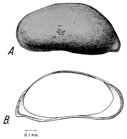
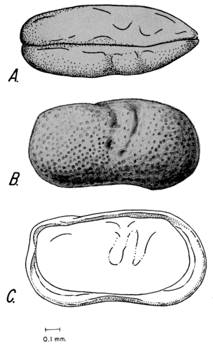
Cypricercus tuberculatus (Sharpe), 1908
Fig. 4. Pl. 2, fig. 11.
Spirocypris tuberculata Sharpe, 1908, Proc. U. S. Natl. Mus., v. 35, p. 406-408, pl. 50, fig. 1-2; pl. 55, fig. 1-6; harpe, 1918, in Ward and Whipple, Fresh-water biology, p. 814, fig. 1267 a-c.
Cypricercus horridus Sars, 1926, Rept. Can. Arctic Exped. 1913-1918, v. 7, pt. 1, p. 6-7; pl. 3, fig. 1-7.
Cypricercus tuberculatus (Sharpe), Hoff, 1942, Illinois Biol. Mono., v. 19, p. 133-135, pl. 8, fig. 103-104.
Diagnosis--The oval shape of the carapace and the papillose surface distinguish this species from the other described species.
Figure 4--Cypricercus tuberculatus (Sharpe). A, Lateral view of exterior of left valve of adult carapace. B, Lateral view of interior of left valve of adult carapace (h, hinge; s, selvage).
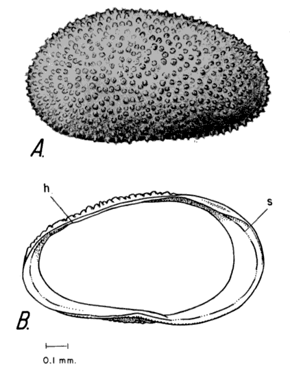
Description--In lateral view the carapace is subelliptical. The dorsal margin is broadly convex. The ventral margin is slightly sinuate in the central ventral region. The anterior margin is broadly rounded. The posterior margin is narrowly rounded. The greatest height is anterior to the middle and exceeds half the length. The surfaces of the valves are covered by numerous papillae. Normal pore canals are numerous and tubelike and extend through the papillae.
In dorsal view the carapace is ovate. The greatest breadth is at the middle and exceeds half the length. The anterior end is more narrowly rounded than the bluntly rounded posterior end. The left valve overlaps the right valve.
The line of concrescence and the inner margin do not coincide, thereby forming a vestibule. The duplicature is widest anteriorly. Radial pore canals are simple, short, numerous, and closely spaced. The marginal flange of the left valve is directed inward. The hingement of the left valve consists of a knifelike bar formed by the hinge flange. The interior surface is pitted; the pits reflect the papillae of the outer surface. The muscle scars could not be determined from the specimens examined.
Males have been recorded for this species, but they cannot be distinguished in fossil specimens.
Measurements--Adult left valve; length 0.91 mm, width 0.48 mm, breadth of whole specimen 0.65 mm.
Remarks--The description and plate diagram of Sars (1926, p. 6, Pl. 3) show that Cypricercus horridus is a synonym of C. tuberculatus. Sharpe (1903) regarded Spirocypris as very close to Cypricercus, differing only in shell shape and size of the furca, which are specific differences. Sharpe (1918) reported the species from springs and shallow weedy and swampy ponds.
Repository--Specimens studied are reposited under catalogue no. 613571.
Genus EUCYPRIS Vávra, 1891
Cypris Müller (part), 1776, Zoologiae Danicae Prodromus, Havniae; Brady and Norman, 1889, Roy. Dublin Soc., Sci. Trans., ser. 2, v. 4, p. 73, 85; Turner, 1895, Rept. Geol. Nat. Hist. Survey Minnesota, ser. 2, ZooL, p. 319; Sharpe, 1898, Bull. Illinois State Lab. Nat. Hist., v. 4, p. 438.
Cypris (Eucypris) Vávra, 1891, Arch. Landesdf. Böhmen, v. 8, pt. 3, p. 82, 84, 90; Alm, 1915, Zool. Bidrag Uppsala, v. 4, p. 46; Furtos, 1933, Ohio Biol. Survey, v. 5 (Bull. 29), p. 450; Dobbin, 1941, Univ. Washington Publ. in Biology, v. 4, no. 3, p. 189-194.
Eucypris Müller, 1912, Das Tierreich, v. 31, p. 168; Sars, 1925, An account of the Crustacea of Norway, v. 9, p. 113; Klie, 1938, Die Tierwelt Deutschlands, v. 34, pt. 3, p. 102-106; Martin, 1940, Senckenbergiana, v. 22, no. 5-6, p. 356; Luttig, 1955, Palaeont. Zeitschr., v. 22, no. 5-6, p. 160.
Type species--Monoculus virens Jurine, 1820.
Diagnosis--This genus is differentiated from the other members of the subfamily Cypridinae by having a strongly arched dorsal margin and by having a sinuation on the central ventral margin. The ventral margin is slightly convex anteriorly. Only in rare individuals is the breadth more than half the length. Upper Cretaceous to Recent.
Description--The carapace is subtriangular in lateral view. The dorsal margin is arched. The posterodorsal and anterodorsal margins form an obtuse angle in the central dorsal region. The ventral margin has a central ventral sinuation and exhibits a small convexity in the anteroventral region. The anterior margin is broadly rounded. The posterior margin is broadly rounded and forms a minor obtuse angle at its junction with the posterodorsal margin. The greatest height is more than half the length. The surface of the valves may be smooth or punctate. Normal pore canals are numerous and simple.
In dorsal view the carapace is ovoid. The greatest breadth equals approximately half the length, and is located posterior to the middle. The anterior end is parabolic. The posterior end is narrowly rounded. The left valve overreaches the right.
In the interior, the line of concrescence and inner margin coincide at the ventral incurvature of the valves. The duplicature is widest anteriorly; the vestibule is very shallow. The hingement is formed by a hinge flange of the left valve and the flange groove of the right valve. The muscle scars are elongate oval and consist of four large scars and one or two smaller scars in a subparallel position.
Remarks--The generic differences in the subfamily Cypridinae have been determined by zoologists on the basis of such internal features as the length of the furcal ramus with respect to its width, features not preserved in fossil carapaces. Eucypris has also been distinguished from Cypricercus because Eucypris reproduces by parthenogensis and males are absent, whereas Cypricercus usually has males. The slightly arched dorsal margin of the carapace of Cypricercus and the more tumid dorsal outline usually differentiate it from Eucypris. The muscle-scar pattern of Cypricercus differs slightly from that of Eucypris. Cyprinotus differs from Eucypris by having tubercles on the margins of the valves.
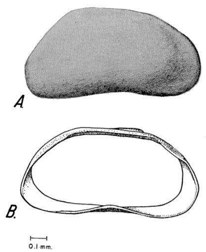
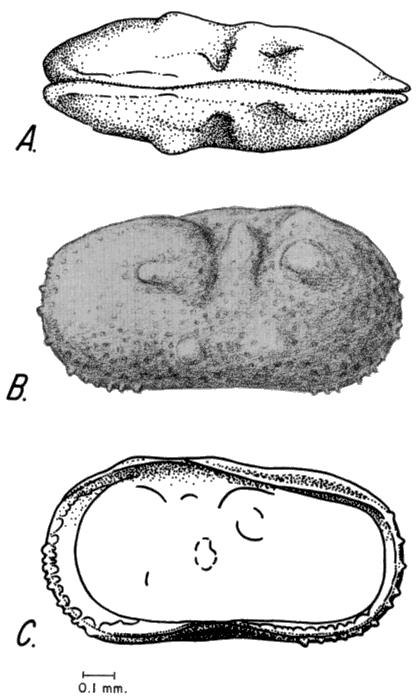
Eucypris Meadensis Gutentag and Benson, n. sp.
Fig. 5. Pl. 2, fig. 8-9.
Diagnosis--The carapace of this species is more compressed and shorter than those of the other, previously described species of Eucypris. The obtuse angle at the junction of the anterodorsal and posterodorsal margins is sharper than in the other species.
Description--Outline of the carapace is subtriangular when seen from lateral view. The dorsal margin is arched in the central dorsal region. The posterodorsal and anterodorsal margins form an obtuse angle in the central dorsal region. The ventral margin has a prominent sinuation in the central ventral region. The ventral margin is slightly convex anterior from the central ventral sinuation. The anterior margin is broadly rounded. The posterior margin is narrowly rounded and forms a small obtuse angle at its junction with the posterodorsal margin. The greatest height is measured from the central dorsal obtuse angle to the ventral margin and is more than half the length. The surface of the carapace is punctate. Normal pore canals are numerous and simple.
In dorsal view, the carapace is elliptical. The greatest breadth is less than half the length, and is located posterior to the middle. The left valve overreaches the right.
Figure 5--Eucypris meadensis n. sp. A, Lateral view of exterior of left valve of adult female carapace. B, Lateral view of interior of left valve of adult female carapace. C, Lateral view of interior of right valve of adult female carapace (d, duplicature; h, hinge groove; 1c, line of concrescence; im, inner margin; rpc, radial pore canals; s, hinge bar; v, vestibule).
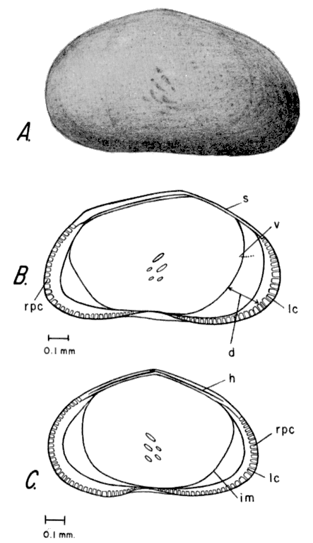
The line of concrescence and the inner margin coincide only at the central ventral incurvature of the valves. The duplicature is widest anteriorly, and present in the posterior; a very narrow vestibule is present. Radial pore canals are moderately long, numerous, and closely spaced. The hingement of the left valve is formed by a knifelike extension of the hinge flange. The posterodorsal hinge flange is directed toward the interior. The hingement of the right valve consists of a groove formed by the hinge flange and the selvage. The flange on the free margin of the left valve is projected inward. A prominent flange groove separates the flange from the slightly raised selvage. The selvage of the right valve is directed upward to fit into the flange groove of the left valve. The muscle scars consist of a subparallel group of five elongate scars whose axes make an angle of about 45° with a tangent to the ventral margin.
Measurements--Holotype: female left valve; length 1.2 mm, height 0.68 mm. Paratype: female right valve; length 1.2 mm, height 0.68 mm, breadth of complete paratype 0.57 mm.
Remarks--This species reproduces only by parthenogenesis. Immature specimens of Eucypris meadensis are more elongate posteriorly in lateral outline than the adults. E. meadensis is found living in an artesian-spring-fed stream in Meade County State Park. Inasmuch as adults were found in the winter as well as the late spring, probably more than one generation is produced each year. The species is associated with Ilyocypris bradyi Sars.
Repository--Specimens studied are reposited under catalogue no. 616571.
Subfamily CYPRIDOPSINAE Kaufmann, 1900
Genus CYPRIDOPSIS Brady, 1868
Cypridopsis Brady, 1868, Intellectual Observer, v. 12, p. 117; Furtos, 1933, Ohio Biol. Survey, v. 5, (Bull. 29), p. 429-430; Dobbin, 1941, Univ. Washington Publ. in Biology, v. 4, p. 230; Swain, 1955, Jour. Paleontology, v. 29, p. 606; Wagner, 1957, The Hague, p. 25.
Pionocypris Brady and Norman, 1896, Roy. Dublin Soc., Sci. Trans., ser. 2, v. 5, p. 725.
Proteocypris Brady, 1906, Nat. Hist. Soc. Northumberland, Trans. ser. 2, v. 1, p. 335.
Pionocypris Sars, 1925, An account of the Crustacea of Norway, v. 9, p. 135.
Type species--Cypris vidua (O. F. Müller), 1776.
Diagnosis--The small size of carapace, distinctive subtriangular shape, and strongly tumid dorsal aspect differentiate Cypridopsis from other members of the subfamily Cypridopsinae. Perm? Upper Cretaceous to Recent.
Description--The carapace is subovoid to subtriangular in lateral view. The dorsal margin is arched. The anterodorsal and posterodorsal margins form a sharp obtuse angle in the central dorsal region. The ventral margin of the right valve is sinuate in the central ventral region. The ventral margin of the left valve is slightly sinuate in some specimens and nearly straight in others. The anterior and posterior margins are broadly rounded. The greatest height, which is near midlength, is much more than half the length. The surfaces of the valves may be smooth or pitted. Normal pore canals are numerous and extend through pits.
In dorsal aspect the carapace is ovate and tumid. The anterior end is pointed; the posterior end is rounded. The breadth is more than half the length. Either valve may be the overreaching valve.
A duplicature with a vestibule is present in the anterior, posterior, and ventral marginal areas. Radial pore canals are not conspicuous. The hinge consists of a bladelike extension of the selvage on the over-reaching valve and a flange groove on the overreached valve. The muscle scar pattern consists of a subcentral group of four large scars and two faint smaller scars. The smaller scars are anterior to the subcentral group.
Remarks--Some workers have assigned species that have the left valve overreaching the right to Pionocypris and have restricted Cypridopsis to those species in which the right valve overreaches the left. The reversal of valve overreach or overlap has been described in other genera, i.e., ? Cytheridea (Alexander, 1933) and is not believed to be of taxonomic importance on the generic level.
Cypridopsis vidua (O. F. Müller), 1776 Fig. 6. Pl. 1, fig. 10.
Cypris vidua O. F. Müller, 1776, Zool. Danicae Prodromus, Havniae, p. 199; Hereick, Ann. Rep. Geol. Nat. Hist. Survey Minnesota, v. 7, p. 112, pl. 18, fig. 1.
Cypridopsis vidua (Müller), Brady, 1868, Intellectual Observer, v. 12, p. 117; Turner, 1892, Bull. Sci. Lab. Denison Univ., v. 6, p. 73; Turner, 1895, Second Rept. Geol. Nat. Hist. Survey Minnesota, p. 312-314, pl. 72, fig. 1-19; pl. 75, fig. 5, 6, 8, 9; pl. 76, fig. 4, 7; Sharpe, 1918, in Ward and Whipple, Fresh-water Biology, p. 807, fig. 1235; Klie, 1938, Die Tierwelt Deutschlands, v. 34, pt. 3, p. 132, fig. 438-441; Hoff, 1942, Illinois Biol. Mono., v. 19, no. 1-2, p. 151-153, pl. 8, fig. 115-117; Bronstein, 1947, Inst. Zool. Acad. Sci. de l'URSS, new ser., no. 31, v. 2, no. 1, p. 160, pl. 9, fig. 8, 10, text fig. 80, 1-3; Kesling, 1951, Illinois Biol. Mono., v. 21, p. 2-116, pl. 1-96, text fig. 1-34; Swain, 1955, Jour. Paleontology, v. 29, p. 606, pl. 60, fig. 6a-c; Wagner, 1957, The Hague, p. 26.
Pionocypris vidua (Müller), Sars, 1925, An account of the Crustacea of Norway, v. 9, p. 135, pl. 63.
Cypridopsis vidua vidua Furtos, 1933, Ohio Biol. Survey, v. 5, (Bull. 29), p. 430-431, pl. 6, fig. 1-4.
Cypridopsis vidua obesa Furtos, 1933, (not Brady and Norman, 1889), Ohio Biol. Survey, v. 5, (Bull. 29), p. 431.
Cypridopsis pustulosa Furtos, 1933, Ohio Biol. Survey, v. 5, (Bull. 29), p. 431-432, pl. 6, fig. 5-9.
Diagnosis--The small size, tumid dorsal appearance, obtuse-angled dorsal margin, and less sinuate ventral margin differentiate this species from the other species of Cypridopsis.
Figure 6--Cypridopsis vidua (O. F. Müller). Lateral view of exterior of right valve of adult carapace.
Description--The carapace of the female is subtriangular in lateral view. The anterodorsal and posterodorsal margins form a sharp obtuse angle on the central dorsal margin. The ventral margin of the right valve is sinuate in the central ventral region. The ventral margin of the left valve is slightly sinuate in some specimens and is nearly straight in other specimens. The anterior margin is more broadly rounded than the posterior margin. The greatest height, which is near midlength, is more than half the length. The surfaces of the valves are pitted. Normal pore canals are numerous and extend through the pits.
In dorsal aspect the carapace is tumid. The anterior end is pointed, the posterior end is broadly rounded. The greatest breadth is in the middle and is much more than half the length. The left valve is slightly larger and overreaches the right valve.
The line of concrescence is slightly removed from the anterior, posterior, and ventral margins. The duplicature is prominent and is widest anteriorly; it is folded in the posteroventral corner in many specimens. Slight upward projection of the free margins of both valves produces left-over-right overlap in the central ventral sinuation. Radial pore canals are short, tubelike, and widely spaced. On most specimens the radial pore canals are inconspicuous. Minute tubercles are found on the anterior margin of the right valve. The hinge is formed by the bladelike extension of the selvage of the left valve into the flange groove of the right valve. At the highest point on the dorsum of the left valve the selvage has a few minute crenulate teeth. On the right valve the corresponding teeth are found outside the selvage, on the selvage groove. On the free margin of the right valve the selvage extends upward and forms a prominent flange groove with the inner margin. Muscle scars consist of a subcentral group of four large scars and two smaller, faint, slightly ventral adductor scars. Two large widely spaced scars are anteroventral to the groups.
Measurements--Adult right valve; length 0.59-0.68 mm, height 0.38-0.44 mm. Adult left valve; length 0.61-0.72 mm, height 0.39-0.49 mm. Adult complete carapace; length 0.63-0.76 mm, breadth 0.47-0.51 mm.
Remarks--Cypridopsis vidua is a multiform species, and recognition of specific differences in the genus is difficult. Probably the differences in the valve surfaces and slight differences in shape can be attributed to variations within the species. C. vidua is well represented in the Pleistocene deposits of Meade County, Kansas. It is also found living in St. Jacobs Well in Clark County, Kansas, where it is associated with other shallow-water genera.
Repository--Specimens studied are reposited under catalogue no. 716561.
Genus POTAMOCYPRIS Brady, 1870
Potamocypris Brady, 1870b, Nat. Hist. Soc. Northumberland and Durham (1869), Trans., v. 3, p. 365; Sharpe, 1898, Bull. Illinois State Lab. Nat. Hist., v. 4, p. 471; Dobbin, 1941, Univ. Washington Publ. in Biology, v. 4, p. 231; Swain, 1955, Jour. Paleontology, v. 29, p. 605.
Candonella Claus, (part) 1891, Arb. Zool. Inst. (Wien), v. 9, p. 231; Vávra, 1898, Ergebn. Hamburg Magalhaensischen Sammelreise, v. 2, p. 12.
Paracypridopsis Kaufmann, 1900, Zool. Anzeiger, v. 23, p. 131.
Cypridopsis (Potamocypris) Alm, 1916, Zool. Bidrag fr. Uppsala, v. 4, p. 83; Furtos, 1933, Ohio Biol. Survey, v. 5, (Bull. 29), p. 432.
Cypridopsella Sars, 1925, An account of the Crustacea of Norway, v. 9, p. 142.
Type species--Bairdia iulva Brady, 1868.
Diagnosis--The arched dorsum, compressed dorsal aspect, and the overreaching right valve differentiate this genus from the other members of the subfamily Cypridopsinae. Upper Cretaceous to Recent.
Description--The carapace is subreniform in lateral view. The dorsal margin is broadly arched. The ventral margin is sinuate in the central ventral region. The anterior margin of the left valve is more broadly rounded than that of the right valve. The posteroventral corner of the left valve in most species is slightly projected posteriorly, where it forms an acute angle. The posterior margin of the right valve is extremely truncate. The greatest height is near midlength and is more than half the length. The surfaces of the valves are smooth in some species but in others may be densely pitted. Normal pore canals are scattered but are most numerous ventrally.
The carapace is elliptical from the dorsal aspect. The anterior end is pointed; the posterior end is rounded. The greatest breadth is much less than half the length. The right valve overreaches the left valve.
In most species the duplicature is widest in the posteroventral corner of the left valve where a prominent flange groove is present. The duplicature of the right valve in most species is welded on the ventral margin. On the rest of the free margin the line of concrescence is removed from the margin and coincides with the inner margin. Radial pore canals are short, tubelike, and closely spaced. The hingement of the right valve consists of a thin groove on the dorsal flange. The hingement of the left valve consists of a bladelike extension of the selvage, which fits into the groove of the right valve. The muscle scars consist of a subcentral group of six oval scars.
Males have been reported for many of the species. Males are more elongate and higher than females.
Remarks--Potamocypris is difficult to identify because of the extreme differences between left and right valves. The genus is common in shallow lakes. The thin valves are usually broken by burial.
Potamocypris smaragdina (Vávra), 1891
Fig. 7. Pl. 1, fig. 4-5.
Cypridopsis smaragdma Vávra, 1891, Arch. naturw. Landesdf. Bohmen, v. 8, pt. 3, p. 80-81, fig. 26; Sharpe, 1898, Bull. Illinois Lab. Nat. Hist., v. 4, p. 470-471, pl. 48, fig. 11-12.
Potomocypris smaragdina (Vávra), Daday, 1900, Ostracoda Hungariae (Budapest), p. 193; Sharpe, 1903, Proc. U. S. Natl. Mus., v. 26, p. 992, pl. 65, fig. 5-7; Sharpe, 1918, in Ward and Whipple, Fresh-water Biology, p. 808, fig. 1245 a-c; Hoff, 1942, Illinois Biol. Mono., v. 19, p. 154-157, pl. 8, fig. 118-124; Swain, 1955, Jour. Paleontology, v. 29, p. 605-606.
Potamocypris smaragdina (Vávra), var. compressa, Furtos, 1933, Ohio Biol. Survey, v. 5, p. 435-437, pl. 6, fig. 10-14; Dobbin, 1941, Univ. Washington Publ. in Biology, v. 4, p. 231-232, pl. 2, fig. 1-6.
Candonella smaragdina Vávra, 1898, Ergebn. Hamburg Megalhaensischen Sammelreise, v. 2, p. 12.
Figure 7--Potomocypris smaragdina (Vávra). A, Lateral view of exterior of left valve of adult carapace. B, Lateral view of exterior of right valve of adult carapace. C, Lateral view of interior of left valve of adult carapace. D, Lateral view of interior of right valve of adult carapace.
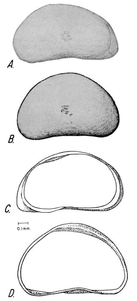
Diagnosis--The shape of P. smaragdina closely approximates that of P. fulva, the type species, but the two species can be distinguished by differences in the adductor muscle-scar pattern. The uppermost scar of P. smaragdina is elongate and complete, whereas that of P. fulva is divided and consists of two separate scars. Other species differences are not distinguishable from examination of the carapace alone.
Description--The adult carapace is elongate subtriangular in lateral view. The anterodorsal and central dorsal margins of the left valve form a rounded obtuse angle anterior to the middle. The dorsal margin of the right valve is broadly arched. The ventral margins of both valves are sinuate in the central ventral region. The anterior margin of the left valve is broadly rounded. The posterior margin of the left valve is extremely truncate and forms an acute angle in the posteroventral corner. The anterior margin of the right valve is more broadly rounded than the posterior margin. The greatest height on the left valve is anterior to the middle and is more than half the length. The greatest height on the right valve is located medially and is more than half to almost two-thirds the length. The valve surfaces are smooth but at high magnification appear slightly roughened. Normal pore canals are scattered over the surfaces of the valves, the greatest concentration being in the ventral half.
Only one complete specimen was available for study. The carapace is elliptical from the dorsal aspect. The posterior end is more bluntly pointed than the anterior end. The greatest width is less than half the length and is located medially. The right valve is larger and overreaches the left valve around the entire margin.
On the right valve the line of concrescence and the inner margin coincide along the free margin. The duplicature is narrow along the anterior margin and is broadest in the posteroventral corner of the left valve. The duplicature is welded along the ventral margin of the right valve but it is absent elsewhere. Radial pore canals are short, tubelike, and widely spaced. They are best observed in the posteroventral area of the left valve. The hinge of the left valve consists of a bladelike valve flange, which fits a groove in the right valve. The anterior and central dorsal valve flange of the right valve is folded toward the interior of the right valve. This is necessary because of the height difference of the valves. The selvage of the left valve along the free margin is projected upward. A prominent flange groove is present in the posteroventral corner of the left valve. The muscle-scar pattern consists of a subcentral group of six, four large scars and two smaller ventral scars.
Males have been reported but they are rare. Hoff (1942, p. 155) stated:
"On the whole, the shell is much more elongate, the height little more than one-half the length; the peak of the dorsal margin more attenuated than in the female."
Measurements--Adult female: left valve, length 0.69-0.74 mm, height 0.38-0.41 mm; right valve, length 0.67-0.72 mm, height 0.41-0.46 mm.
Remarks--Potamocypris smaragdina is difficult to describe because of intraspecific variation in its size and shape. It is found in the Pleistocene deposits of Meade County. Hoff (p. 156) reports P. smaragdina as a common species of permanent waters.
Repository--Specimens studied are reposited under catalogue no. 716561.
Family CANDONIDAE Kaufmann, 1900
Genus CANDONA Baird, 1846
Candona Baird, 1846a, p. 152; Brady, 1868, Linnean Soc. London Trans., v. 26, p. 381; Brady and Norman, 1889, Royal Dublin Soc., Sci. Trans., ser. 2, v. 4, p. 98; Kaufmann, 1900, Revue Suisse Zool., v. 4, p. 379; Müller, 1900, Zoologica, v. 30, p. 15; Klie, 1938, Die Tierwelt Deutschlands, v. 34, pt. 3, p. 25-26; Swain, 1947, Jour. Paleontology, v. 21, p. 520-521; Triebel, 1949, Senckenbergiana, v. 30, no. 4-6, p. 205-212; Swain, 1955, Jour. Paleontology, v. 29, p. 607-609; Luttig, 1955, Palaeont. Zeitschrift, v. 29, no. 3-4, p. 151; Wagner, 1957, The Hague, p. 18.
Eucandona Daday, 1900, Ostracoda Hungariae (Budapest), p. 242; Swain, 1961, Treatise on Invert. Paleontology, pt. Q, Arthropoda 3, p. Q 234-235.
Type species--Cypris Candida Müller, O. F., 1776.
Diagnosis--The reniform shape of the carapace and the muscle-scar pattern, a subcentral rosette of five scars and one dorsal elongate scar, are features that differentiate Candona from the other genera in the Family Candonidae. Paleogene to Recent.
Description--In lateral view, the carapaces of both sexes are nearly reniform. The dorsal margin is approximately straight to broadly curved. The ventral margin is approximately straight to concave. The anterior margin is rounded; the posterior margin is rounded in the males, more truncate than the anterior margin in the female. The greatest height is in the middle or posterior to the middle. Surfaces of the valves are smooth or minutely punctate, and reticulate in some species. Normal pore canals are simple and scattered over the surfaces of the valves.
From the dorsal aspect the carapace is sub-elliptical. The anterior end is pointed, but the posterior end is rounded. The left valve overreaches the right valve.
The line of concrescence and the inner margin seem to coincide only at the central ventral incurvature of species that have sinuations. The duplicature is widest in the anterior end. Radial pore canals are numerous, simple, and closely spaced. A hinge flange on the selvage of the right valve fits into a groove on the selvage of the left valve. Muscle scar pattern consists of a subcentral rosette of five scars and one dorsal elongate scar.
Sexual dimorphism is pronounced in some species. The concave ventral margin of the male is distinctive, and in many species the anteroventral margin meets the ventral sinuation of a sharp acute angle. The males of many species are more inflated than the females.
Remarks--Howe (1955) discussed the discovery of a paper by Baird (1846b) in which Candona reptans was designated as the type for Candona. C. reptans was designated by Brady and Norman in 1889 as the type species for Erpetocypris, and C. Candida was designated as the type for Candona. Swain (1961) assigned all the forms that were previously placed in Candona to Eucandona and designated C. balatonica as the type species. According to the action taken by the International Commission of Zoological Nomenclature (1958, opinion 533), through application of its plenary powers, Cypris Candida Müller was designated as the type species of Candona; Eucandona then became a junior subjective synonym.
Recent species inhabit fresh-water lakes and ponds and brackish bays. They are creepers and burrowers instead of swimmers. They are holarctic in distribution, and in this environment they are present in the largest number of species of any genus in the Cypridacea.
Candona caudata Kaufmann, 1900
Fig. 8. Pl. 2, fig. 11.
Candona caudata Kaufmann, 1900. Revue Suisse Zool., v. 8, p. 367-368, pl. 24, fig. 16-20; pl. 26, fig. 17-23; Vávra, 1909, Die Süsswasser Fauna Deutsch., v. 2, p. 96, fig. 391-392; Alm, 1915, Zool. Bidrag. Uppsala, v. 4, p. 190; Sars, 1925, An account of the Crustacea of Norway, v. 9, p. 76-77, pl. 35; Klie, 1938, Die Tierwelt Deutschlands, v. 34, p. 68, pl. 3, fig. 223-225; Hoff, 1942, Illinois Biol. Mono., v. 19, p. 80-82, pl. 3, fig. 33-35; Swain, 1955, Jour. Paleontology, v. 29, p. 608, pl. 29, fig. 5.
Candona elongata Müller, 1912 (non Herrick, 1879), Das Tierreich, v. 31, p. 140.
Diagnosis--The posterior margin of the female left valve has a distinctive hook-shaped, acutely angled extension at the junction of the posterior and ventral margins. The right valve has a smaller extension in the posteroventral corner.
Description--In lateral view, the carapace is elongate and reniform. The dorsal margins of both valves are arched, but the right valve has a small sinuation in the anterodorsal region. The ventral margin is sinuous in the central ventral region. The left valve is also slightly sinuous on the posteroventral margin. The anterior margins of both valves are bluntly rounded. The posterior margin of the right valve is sharply rounded and has a small extension in the posteroventral extremity. The posterior margin of the left valve has a sharply acute angled, hook-shaped posteroventral extension, which curves toward the right valve. The greatest height is posterior to the middle and is" less than half the length. The surface of the valves is smooth and translucent; many specimens have been diagenetically bleached.
Figure 8--Candona caudata Kaufmann. A, Lateral view of exterior of left valve of adult female carapace. B, Lateral view of interior of left valve of adult female carapace.
Only separated valves were available for study, but the relative shape of the valves clearly shows that the left overreaches the right.
In the interior view, the line of concrescence and the inner margin are widely separated except at the central ventral infolding and the dorsal margin. The duplicature is widest anteriorly and in the posteroventral areas. Radial pore canals are numerous, short, and closely spaced. The hinge consists of a groove on the dorsum of the left valve, which accommodates the bladelike edge of the right valve. The muscle scars are similar to those described for the genus.
Measurements--Female left valve; length 1.17 mm, height 0.57 mm. Female right valve; length 1.12 mm, height 0.54 mm.
Remarks--Candona caudata Kaufmann is distinguished from C. crogmaniana Turner by the hook-shaped extension on the left valve. C. crogmaniana is also larger and more truncate posteriorly than C. caudata.
Repository--Material studied is reposited under catalogue no. 927567.
Candona Crogmaniana Turner, 1894
Fig. 9. Pl. 2, fig. 10.
Candona crogmaniana Turner, 1894, Bull. Sci. Lab. Denison Univ., v. 8, p. 20-21, pl. 8, fig. 24-33; Furtos, 1933, Ohio Biol. Survey, v. 5, (Bull. 29), p. 476, pl. 8, fig. 1-3; pl. 9, fig. 17-18; pl. 12, fig. 9-10; Hoff, 1942, Illinois Biol. Mono., v. 19, p. 79-80, pl. 3, fig. 31-32.
Candona crogmani Turner, 1895, Rept. Geol. Nat. Hist. Survey Minnesota, ser. 2, Zool., p. 300-301, pl. 71, fig. 24-33, pl. 81, fig. 405; Sharpe, 1918, in Ward and Whipple, Freshwater Biology, p. 824, fig. 1295a-c.
Diagnosis--The subtriangular carapace, the strongly arched dorsal margin, and the truncate posterior end distinguish this species from the other members of the genus.
Figure 9--Candona crogmaniana Turner. A, Lateral view of exterior of right valve of adult female carapace. B, Lateral view of interior of left valve of adult female carapace.
Description--In lateral view, the carapace is elongate and subtriangular. The dorsal margin is strongly arched posteriorly. The right valve has a small sinuation in the anterodorsal region. The central dorsal margin is straight; the ventral margin is concave. The anterior margin is narrowly rounded. The posterior margin is truncate forming an acute angle of about 53° with the venter. The greatest height, in the posterior end, is almost half the length. The surface of the valves is smooth. Normal pore canals are simple and scattered at random over the surface.
Only a few right valves of the female were available for study. The dorsal view (Furtos, 1933, p. 476) is as follows:
"Compressed, elliptical, over two and one-half times longer than broad; extremities pointed, the posterior the narrower; left valve longer than the right, projecting considerably beyond it at the posterior extremity."
In interior view, the line of concrescence is slightly removed from the ventral, posteroventral, and anterior margins. The duplicature is widest in the anterior end. The radial pore canals are short, closely spaced, and very numerous. The hinge consists of a groove on the dorsum of the left valve that accommodates a bar on the dorsum of the right valve. The muscle scars are similar to those described for the genus.
Measurements--Female right valve; length 1.18 mm, height 0.65 mm.
Remarks--The female of Candona crogmaniana Turner is similar to that of C. caudata Kaufmann but the former has a higher arch and a less pronounced posteroventral angle. The earliest occurrence of C. crogmaniana is from the Laverne Formation (Lower Pliocene). It is comparatively rare but its distinctive features make it readily identifiable when present.
Repository--Specimens studied are reposited under catalogue no. 717562.
Candona fluviatilis Hoff, 1942
PI. 1, fig. 13
Candona fluviatilis Hoff, 1942, Illinois Biol. Mono., v. 19, p. 60-62, pl. 1, fig. 7-9; pl. 2, fig. 10-11.
Diagnosis--The reticulate surface and trapezoidal lateral outline distinguish this species from the other members of the genus.
Description--The carapace of the female is trapezoidal in lateral view. The anterodorsal margin is slightly sinuous. The anterodorsal and central dorsal margins form a distinct obtuse angle. The central dorsal margin is nearly straight, only faintly concave. The central dorsal margin and the posterodorsal margin form a broadly rounded obtuse angle. The posterodorsal margin is broadly convex. The central ventral margin is sinuate, the greatest concavity being slightly anterior to the middle. The anterior margin is broadly rounded. The posterior margin is truncate and forms a narrpwiy rounded posteroventral corner. The greatest height is posterior to the middle and is slightly more than half the length. The surface of the valve is sub-reticulate. The reticulae are composed of interconnected ridges that surround small polygonal depressions. Normal pore canals are not visible.
Only a female left valve was available for identification; therefore, a description of the dorsal view is omitted. The structure of the hinge indicates that the left valve overreaches the right valve.
Inspection of the interior of the left valve discloses a fused duplicature widest at the anterior and present in the posterior. Radial pore canals are short, simple, and closely spaced. The hinge of the left valve consists of a groove in the dorsum formed by the hinge flange and a bladelike extension of the selvage. Muscle scars are the same as those described for the genus.
Measurements--Adult female; length 0.88 mm, height 0.45 mm.
Remarks--Candona fluviatilis Hoff is distinguished from C. punctata Furtos by its straight dorsal margin, reticulate surface, and smaller height. Hoff (1942, p. 62) collected this species from clear, shallow vernal streams flowing over muddy bottoms.
Repository--Specimen studied is reposited under catalogue no. 927567.
Candona nyensis Gutentag and Benson, n. sp.
Fig. 10. Pl. 2, fig. 1-3.
Candona sp. aff. Cypris pubera O. F. Müller, Swain, 1947, Jour. Paleontology, v. 21, pl. 76, fig. 14-16.
Fig. 10--Candona nyensis n. sp. A, Lateral view of exterior of right valve of adult male carapace. B, Lateral view of exterior of left valve of adult female carapace. C, Lateral view of interior of left valve of adult female carapace. D, Lateral view of interior of left valve of adult male carapace. (ce, caudal extension; d duplicature; dm, dorsal margin; h hinge grove; hf, hinge flange; im, inner margin; lc, line of concrescence; om, outer margin; rpc, radial pore canals.)
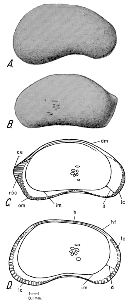
Diagnosis--The extremely truncate posterior margin of the female left valve and the obtuse anteroventral angle of the male carapace distinguish this species.
Description--The female in lateral view is subreniform. The dorsal margin of the left valve is arched and there is a small sinuation in the posterodorsal region. The ventral margin is concave. The anterior margin is narrowly rounded. On the right valve, the posterior margin is bluntly rounded where the posterior and ventral margins form an obtuse angle. On the left valve, the posterior margin is extremely truncate. Above midheight, dorsal margin forms an obtuse angle with the posterior margin (posterior cardinal angle). The posterior margin is almost vertical as it slopes away from the posterodorsal angle, and it forms an obtuse angle with the ventral margin (posteroventral angle). The greatest height is posterior to the middle of the valve and is more than half the length. The surface of the valves is smooth and the shell material of many specimens is translucent. Many simple normal pore canals are scattered throughout, the greatest concentration being in the ventral region.
In dorsal view, the female carapace is elliptical in outline. The anterior end is pointed, the posterior end is truncate, and the posterior margin of the left valve does not touch the right valve at the posteroventral angle. The left valve overreaches the right.
In the interior view, the line of concrescence is submarginal except at the incurvature on the ventral margin, where it is missing. The duplicature is widest anteriorly. Radial pore canals are simple, numerous, and closely spaced; they are not present at the infolding of the valves in the central ventral region. The hingement consists of a thin flange and grooved selvage on the left valve and a flange and thin selvage on the right valve. The muscle scars consist of a group of five scars as described for the genus.
Sexual dimorphism is pronounced in this species. The carapace of the male in lateral view is arched and larger than the female. The dorsal margin is strongly arched at the posterior termination of the hinge. The anterior is rounded; the posterior end is broadly rounded. The ventral margin is strongly sinuate, the greatest concavity being in the anterior end. The anteroventral angle is obtuse and is formed by the ventral sinuation and the anteroventral margin.
Measurements--Holotype, female left valve; length 1.28 mm, height 0.74 mm. Paratype, female right valve; length 1.18 mm, height 0.65 mm. Allotype, male left valve; length 1.3 mm, height 0.80 mm. Allotype, male right valve; length 1.05 mm, height 0.85 mm.
Remarks--This species was illustrated by Swain (1947, pl. 76, fig. 14-16 as Candona sp. off. Cypris pubera O. F. Müller. Swain found Candona nyensis in the Recent deposits of Lake St. John, Colorado. C. nyensis was described by Staplin (1953, p. 162-166) as "Candona swaini." This species seems to be a dominantly Pleistocene form, possible relics still living in cold-water stable habitats.
Repository--Specimens studied are reposited under catalogue no. 716561.
Candona renoensis Gutentag and Benson, n. sp.
Fig. 11. Pl. 1, fig. 12.
Fig. 11--Candona renoensis n. sp. A, Lateral view of exterior of left valve of adult female carapace. B, Lateral view of exterior of left valve of adult male carapace. C, Lateral view of interior of left valve of adult female carapace. D, Lateral view of interior of left valve of adult male carapace. (d duplicature; g hinge grove; hf, hinge flange; lc, line of concrescence; im, inner margin; rpc, radial pore canals; v, vestibules.)
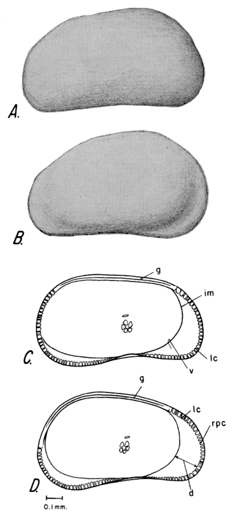
Diagnosis--The subtrapezoidal lateral margin and the compressed female carapace clearly distinguish this species from those previously described. The male carapace has a distinctive pointed anterior end and is more strongly inflated than the female.
Description--The carapace of the female is subtrapezoidal and reniform in lateral view. A slight sinuation is present on the anterodorsal margin. The central dorsal margin is nearly straight. A broadly rounded obtuse angle is formed by the central dorsal and posterodorsal margins. The ventral margin is slightly sinuous in the central ventral area. The anterior margin is broadly rounded. The posterior margin is truncate. The posteroventral corner is a rounded obtuse angle. The greatest height is posterior to the middle and is slightly more than half the length. The surfaces of the valves are smooth, and the valves are translucent. Normal pore canals are simple and scattered over the surface, the greatest concentration being in the ventral area.
In dorsal view, the carapace of the female is narrow and elliptical. The anterior end is narrowly rounded. The posterior end is bluntly rounded. The left valve overreaches the right valve slightly, completely around the margin.
The marginal area is simple, composed of a wide anterior and narrower posteroventral duplicature forming a vestibule. The duplicature is absent or almost so from the posterior margin. Radial pore canals are numerous, short, and simple. The hingement consists of a hinge flange on the right valve that fits into the groove on the left valve. The groove on the left valve is formed by the hinge flange and a bladelike extension of the selvage. The muscle scar pattern is like that typical of the genus.
Sexual dimorphism is pronounced. The carapace of the male is more arched than the female; in dorsal view the anterior end is narrow and sharp because of the severe flattening of the anterior margin. The posterior end is broadly rounded. The breadth of the male is much greater than that of the female, which gives the male an inflated ovate shape. The marginal features of the male are the same as those of the female.
Measurements--Holotype: left valve of female; length 0.99 mm, height 0.53 mm. Paratype: right valve of female; length 0.94 mm, height 0.51 mm. Breadth of female paratype 0.35 mm. Allotype: left valve of male; length 1.01 mm, height 0.61 mm; breadth of allotype 0.45 mm.
Remarks--Candona renoensis differs from Candona compressa Koch by the more compressed breadth of the female. It differs from Candona albicans Brady by having a more rounded posterodorsal margin. The male of C. renoensis has a narrowly rounded anterior, which swells into an inflated carapace and it is therefore distinctive.
Repository--Specimens studied are reposited under catalogue no. 613571.
Family ILYOCYPRIDIDAE Kaufmann, 1900
Subfamily ILYOCYPRIDINAE Kaufmann, 1900
Genus ILYOCYPRIS Brady and Norman, 1889
Ilyocypris Brady and Norman, 1889, Roy. Dublin Soc., Sci. Trans. ser. 2, v. 4, p. 106; Furtos, 1933, Ohio Biol. Survey, v. 5, (Bull. 29), p. 426; Klie, 1938, Die Tierwelt Deutschlands, v. 34, pt. 3, p. 90; Hoff, 1942, Illinois Biol. Mono., v. 19, p. 127-128; Triebel, 1941, Senckenbergiana, v. 23, no. 4-6, p. 298; Luttig, 1955, Palaeont. Zeitschr., v. 29, no. 3/4, p. 161.
Iliocyprella Daday, 1900, Ostracoda Hungariae, p. 237.
Type species--Cypris gibba Ramdohr, 1808.
Diagnosis--The carapace is nearly oblong but has a straight dorsal margin. One or two dorsal lateral sulci are present. Lateral protrusions occur in many species. Triassic to Recent.
Description--The carapace is nearly oblong in the laterial view. The dorsal margin is almost straight. The ventral margin is sinuate in the central ventral region. The anterior margin is more broadly rounded than the posterior, and the anterior sulcus is larger than the posterior. The greatest height is anterior to the center. The surface of the valves is reticulate. Pits, nodes, and spines may be present. In most species spines are present along the entire anterior, ventral, and posterior margins. Larger spines are present in the posterior region.
As seen from the dorsal view most species have two or three lateral protrusions. The anterior end is narrower than the posterior, and the left valve is slightly larger than the right.
The line of concrescence in most forms is removed from the ventral, posteroventral, and anterior margins. The duplicature is widest anteriorly and in the posteroventral area. Radial pore canals are simple and very numerous but may be obscured by the ornamentation. The hinge consists of a groove in the left valve into which fits the bladelike dorsal edge of the right valve. A rosette of four muscle scars is situated in a subcentral depression.
Ilyocypris bradyi Sars, 1890
Fig. 12. Pl. 1, fig. 8-9.
Ilyocypris bradyi Sars, 1890, Oversigt af Norges Crustaceer, v. 2, p. 59-60; Sharpe, 1908, Proc. U. S. Natl. Mus., v. 26, p. 411-412, pl. 56, fig. 3-6; Sharpe, 1918, in Ward and Whipple, Fresh-water Biology, p. 810, fig. 1258 a-d; Klie, 1938, Die Tierwelt Deutschlands, v. 34, pt. 3, p. 93, fig. 329-332; Hoff, 1942, Illinois Biol. Mono., v. 19, p. 130-131, pl. 8, fig. 101-102.
Ilyocypris bradii Furtos, 1933, Ohio Biol. Survey, v. 5, (Bull. 29), p. 428, pl. 1, fig. 8-10.
Figure 12--Ilyocypris bradyi Sars. A, Dorsal view of joined valves of adult carapace. B, Lateral view of exterior of right valve of adult carapace. C, Interior view of left valve of adult carapace.
Diagnosis--Lack of lateral protrusions and the presence of two distinct dorsolateral sulci on Ilyocypris bradyi distinguish it from other species of the genus.
Description--Carapace in lateral view is subquadrate in outline. The dorsal margin is approximately straight. The anterior sulcus is larger than the posterior sulcus. A median lobe separates the sulci. The ventral margin is sinuate in the central ventral region. The anterior margin is broadly rounded; the posterior margin is narrower but more bluntly rounded. The greatest height is anterior to the center. The surface of the valves is reticulate; numerous spines project from anterior, posterior, and ventral margins. The spines along the posterior margin are larger than the others. In some specimens tubercles are present along all margins except the dorsal.
In dorsal view, the outline of the carapace is narrowly elliptical. The anterior end is pointed. The posterior end is narrowly rounded and slightly inflated. The dorsal surface is depressed at the point of contact of the valves between the median lobe and the posterior inflated area. The hinge flange of both valves projects slightly at the anterodorsal margin. The left valve overreaches the right.
The line of concrescence is removed from the ventral, posteroventral, and anterior margins. The duplicature is widest anteriorly. Radial pore canals are simple and are not regularly spaced. On the left valve the hinge consists of a groove, which is best seen in the anterodorsal corner. On the right valve the hinge consists of a knife-like edge, which fits the groove on the left valve. The muscle-scar pattern consists of a group of four scars in a pit in the subcentral area.
Males are not known for this species.
Measurements--Adult female: length 0.80-0.91 mm, height 0.42-0.47 mm, breadth 0.31-0.40 mm.
Remarks--Ilyocypris bradyi lives in moving water where there is an abundance of algae and other vegetation. It was found living in association with Eucypris meadensis in the swift-flowing artesian-spring-fed stream in Meade County State Park, Kansas. I. bradyi does not compete with E. meadensis, as the former is a burrowing organism and the latter is a bottom crawler.
Repository--Specimens studied are deposited under catalogue no. 927567.
Ilyocypris gibba (Ramdohr), 1808
Fig. 13. Pl. 1, fig. 6-7.
Ilyocypris gibbo (Ramdohr), Brady and Norman, 1889, Roy. Dublin Soc., Sci. Trans., ser. 2, v. 4, p. 107, pl. 22, fig. 1-5; Sharpe, 1908, Proc. U. S. Natl. Mus., v. 35, p. 410-411, pl. 56, fig. 1-2. Sharpe, 1918, in Ward and Whipple, Fresh-water Biology, p. 809, fig. 1257a,b; Furtos, 1933, Ohio Biol. Survey, v. 5, (Bull. 29), p. 427, pl. 1, fig. 4-7; Klie, 1938, Die Tierwelt Deutschlands, v. 34, pt. 3, p. 90, fig. 316; Hoff, 1942, Illinois Biol. Mono., v. 19, p. 128-130, pl. 7, fig. 99-100; Bronstein, 1947, Inst. Zool. Acad. Sci. de l'URSS, new ser., no. 31, v. 2, no. 1, p. 88, pl. 1, fig. 5-6; Luttig, 1955, Palaeont. Zeitschr. v. 29, no. 3-4, p. 161, pl. 17, fig. 5-9.
Figure 13--Ilyocypris gibba (Ramdohr). A, Dorsal view of joined valves of adult carapace. B, Lateral view of exterior of right valve of adult carapace. C, Lateral view of interior of right valve of adult carapace.
Diagnosis--The presence of two distinct dorsolateral sulci, three prominent lateral protrusions, and numerous nodes distinguish this species from the other members of the genus.
Description--The adult female, in lateral view, is nearly oblong. The dorsal margin is nearly straight but is slightly arched over the sulci and it appears depressed posteriorly where the posterior lobe projects above the hinge line. The two sulci are broadest at the dorsal margin but they narrow considerably toward the venter and terminate in the dorsocentral area. The anterior sulcus is longer than the posterior sulcus. The ventral portion of the anterior sulcus is longer than the ventral portion of the posterior sulcus. The ventral margin is sinuate in the central ventral region. The anterior margin is more broadly rounded than the posterior margin. The greatest height is in the anterior and exceeds half the length. The surfaces of the valves are pitted. A median lobe separates the sulci and bears a small node. The anterior lobe bears a small node just anterior to the sulcus. A large node in the dorsocentral region is posterior to the termination of the posterior sulcus. Many small spines and tubercles are scattered along the submarginal and marginal areas. Normal pore canals are inconspicuous because of the ornamentation.
The carapace outline is elliptical in dorsal view. The anterior end is pointed. The posterior end is narrowly rounded. The contact margin appears depressed along the posterior half where the two posterior lobes bulge dorsally. The breadth is less than half the length. The left valve is larger and overreaches the right valve.
The duplicature is widest anteriorly and has a small groove along the anterior part and a slight fold near the inner margin. Radial pore canals are simple and numerous. On the left valve the hinge consists of a groove, which is more pronounced in the anterodorsal and posterodorsal corners. On the right valve the hinge consists of a simple blade-like edge, which fits the groove in the left valve. The muscle-scar pattern consists of four scars in a subcentral pit below the posterior sulcus.
Measurements--Adult female; left valves, length 0.80-0.95 mm, height 0.44-0.52 mm; right valves, length 0.80-0.96 mm, height 0.43-0.53 mm. Breadth of complete carapace 0.35 mm, length 0.84 mm.
Remarks--Ilyocypris gibba is very common in running water but is also found in ponds and lakes containing algae and other vegetation. Lüttig (1955) reports that I. gibba is found in the Recent of Europe, North Africa, and North America, and in the Quaternary of Great Britain. Klie (1938) states that I. gibba does not live in water colder than 50 °F.
Repository--Specimens studied are reposited under catalogue no. 61357.
Superfamily CYTHERACEA Baird, 1850
Family CYTHERIDEIDAE Sars, 1925
Subfamily CYTHERIDEINAE Sars, 1925
Genus CYPRIDEIS Jones, 1856
Cyprideis Jones, 1857, Palaeontographical Soc. London (1856), v. 4, p. 21; Sars, 1925, An account of the Crustacea of Norway, v. 9, p. 154; Bronstein, 1947, Inst. Zool. Sci. de l'URSS, new ser., no. 31, v. 2, no. 1, p. 295; Goerlich, 1952, Senckenbergiana, v. 33, no. 1-3, p. 185-186; Swain, 1955, Jour. Paleontology, v. 29, p. 614.
Anomocytheridea Stephenson, 1938, Jour. Paleontology, v. 12, p. 141.
Type species--Candona torosa JONES, 1850.
Diagnosis--This genus differs from the other members of the sub-family Cytherideinae by its reniform to trapezoidal outline and by the occurrence of pits and occasional tubercles on the surface. Sexual dimorphism is pronounced in the genus. Miocene to Recent.
Description--The female carapace, in the lateral view, is reniform to trapezoidal. The whole dorsal margin in some species is nearly straight or slightly convex, but it may slope toward the posterior. The ventral margin is slightly sinuate in most species but may be nearly straight in others. The anterior margin is broadly rounded. The posterior margin is truncate and in most species meets the venter in a rounded or sharp acute angle. The greatest height is anterior to the middle and is equal to or more than half the length. Normal pore canals are located in the pits. The surfaces of the valves are pitted; tubercles may be present on some species. Within some species, nodes are present or absent in the younger instars. Many species have a dorsal lateral depression.
The carapace in dorsal view is subelliptical. The anterior end is narrowly rounded, the posterior bluntly rounded. The left valve overreaches the right.
The line of concrescence coincides with the inner margin on the anterior and posterior margins. The duplicature is normally welded, but in some species a thin free duplicature is found inside the ventral margin. Numerous radial pore canals are present. They are tubelike and some may bifurcate near the outer margin. They form groups of two or three, and the groups are slightly separated from one another. The hinge of the right valve consists of an anterior crenulated tooth, a tooth socket, and a posterior tooth separated from the anterior tooth socket by a thin crenulated groove. The left valve is in antithesis to the right valve. The muscle scars consist of a row of four closely spaced adductor scars anterior to the middle but in the central area, and two widely spaced anterior scars.
Sexual dimorphism is pronounced; the posteroventer is generally extended in males. The males are larger than the females in all described species.
Remarks--Goerlich (1952, p. 186) shows that the hinge structure of Anomocytheridea as described by Stephenson is the same as that of Cyprideis; therefore Anomocytheridea is a synonym of Cyprideis. Cyprideis has been reported from Miocene to Recent and it is euryhaline, occurring in marine, brackish, and fresh water.
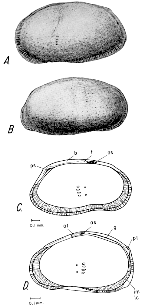
Cyprideis littoralis Brady, 1870
Fig. 14. Pl. 2, fig. 4-7.
Cyprideis littoralis Brady, 1870a, Nat. Hist. Soc. Northumberland and Durham, Trans., v. 3, p. 125; Sars, 1925, An account of the Crustacea of Norway, v. 9, p. 155. pl. 71, pl. 72, fig. 1; Klie, 1938, Die Tierwelt Deutschlands, v. 34, pt. 3, p. 156. fig. 516-517; Bronstein, 1947, Inst. Zool. Acad. Sci. de l'URSS, new ser., no. 31, v. 2, no. 1, p. 296-297, pl. 14, fig. 6-7; Swain, 1955, Jour. Paleontology, v. 29, p. 615-616, pl. 59, fig. 11 a-c, text-fig. 38, 5 a, b.
Cytheridea torosa littoralis (Brady) Müller, 1912, Das Tierreich, v. 31, Ostracoda, p. 326.
Cytheridea torosa vax. teres Brady and Norman, 1889, Royal Dublin Soc., Sci. Trans., v. 4, ser. 2, p. 175.
Figure 14--Cyprideis littoralis Brady. A, Lateral view of exterior of left valve of adult male carapace. B, Lateral view of exterior of right valve of adult male carapace. C, Lateral view of interior of left valve of adult male carapace. D, Lateral view of interior of right valve of adult female carapace. (as, anterior tooth socket; at, anterior tooth; b bar; g, groove; im, inner margin; lc, line of concrescence; ps, posterior hinge socket; pt, posterior hinge tooth; t, anterior hinge tooth.)
Diagnosis--The extremely truncate posterior margin of the female, the muscle-scar pattern (a row of four small round adductor scars), and the dorsolateral sulcus distinguish this species from other members of the genus.
Description--Female carapace is subovate in lateral view. The dorsal margin of the right valve is slightly more convex than that of the left valve. The anterodorsal margin is slightly sinuate. A slightly rounded obtuse angle is formed at the junction of the ventral and posterior margins. The anterior margin is broadly rounded. On the right valve a submarginal extension of the selvage is exposed along the anterior margin. The posterior margin is extremely truncate--almost vertical. The greatest height is located medially and is more than half the length. The surfaces of the valves are pitted. A shallow elongate dorsal sulcus is located anterior to the middle. Normal pore canals are numerous and extend from the interior through the pits.
In dorsal view the female carapace is subelliptical. The anterior is pointed; the posterior is bluntly rounded. The dorsal lateral sulci are slightly anterior to the middle. The hinge flange and the selvage flange project upward along the dorsal margin and have an exposed groove. The left valve is slightly larger and overreaches the right valve.
The line of concrescence coincides with the inner margin along the anterior and posterior margins of the valves. A narrow duplicature is present along the ventral margin posterior to the middle and extending to the posteroventral corner. Radial pore canals are numerous long tubes, which may show some bifurcation, and most are found in separate pairs. The hinge of the right valve consists of a raised crenulate dental element on the raised selvage extension anterior to the middle on the dorsal margin. An intermediate groove posterior to the raised crenulate anterior dental element is followed by a triangular tooth socket. Posterior to the tooth socket a crenulate groove is formed by the selvage and the valve edge. This thin groove extends from the tooth socket to the posterodorsal corner, where the valve edge is raised to form a crenulate posterior tooth. The hinge of the left valve is in antithesis to that of the right valve. The blade-like selvage of the free margin of the right valve fits the thin groove formed by the selvage and valve edge on the left valve. The muscle-scar pattern consists of a row of four small round adductor scars anterior to the middle but in the central region. Two widely spaced antennal and mandibular scars are located anterior to the row of adductor scars.
Sexual dimorphism is pronounced in this species. The carapace of the male is larger and more elongate than that of the female. The dorsal margin is evenly arched. The posterior margin is broadly rounded. The posteroventral acute angle is sharper than in the female. The features of the interior of the male valve are the same as those in the female.
Measurements--Adult female: left and right valves; length 0.99-1.03 mm, height 0.63 mm, breadth 0.99-1.0 mm. Adult male: left valve; length 1.18-1.22 mm, height 0.67-0.69 mm; right valve; length 1.19-1.23 mm, height 0.65-0.67 mm.
Remarks--Cyprideis littoralis is found in the Laverne Formation (Pliocene) and in fresh-water Pleistocene sink deposits in Meade County, Kansas. C. littoralis seems to be euryhaline. Swain (1955, p. 616) reports this species from the middle and lower parts of San Antonio Bay, Texas, and in nearshore or brackish waters in Europe, Asia, and northern Africa. The measurements of C. littoralis from the Pleistocene of Meade County compare closely to those given by Sars (1925, p. 155). On the Pleistocene specimens the dorsal margin seems slightly more arched than that on the European specimens.
Repository--Specimens studied are reposited under catalogue no. 716561.
Family LIMNOCYTHERIDAE Klie, 1938
Genus LIMNOCYTHERE Brady, 1868
Limnicythere Brady, 1868, Intellectual Observer, v. 12, p. 121; Klie, 1938, Die Tierwelt Deutschlands, v. 34, pt. 3, p. 150; Luttig, 1955, Palaeont. Zeitschrift, v. 29, no. 3-4, p. 162; Swain, 1955, Jour. Paleontology, vol. 29, p. 612.
Limnicythere Brady, 1868, Linnean Soc., Trans., (London), v. 26, p. 419; Sars, 1925, An account of the Crustacea of Norway, v. 9, p. 149; Furtos, 1933, Ohio Biol. Survey, v. 5, (Bull. 29), p. 422; Dobbin, 1941, Univ. Washington Publ. in Biology, v. 4, no. 3, p. 185.
Type species--Cythere inopinata Baird, 1843.
Diagnosis--The subrectangular shape of the carapace and the distinctive surface ornamentation and hingement of both sexes differentiate this genus from the other members of the family Limnocytheridae. Jurassic to Recent.
Description--Carapace is subreniform to subrectangular in lateral view. In most species the dorsal margin is straight, but it may be slightly arched in some species. The ventral margin is sinuate; in most species the greatest concavity is in the central ventral region. The anterior margin is broadly rounded. The posterior margin is narrowly rounded. The greatest height is anterior to the middle in most species, and is approximately half the length. The surfaces of the valves are pitted or reticulate in most species. One or two transverse sulci, prominent along the dorsal margin, fade ventrally and disappear above the ventral margin. Nodes occur in many species. The marginal areas are flattened and may have small tubercles or spines.
In dorsal view, the carapace is compressed. The anterior is sharply pointed; the posterior is narrowly rounded. The dorsal valve flanges project upward. The sulci are very distinct. Valves of most species are inflated in the middle. Slight alate lateral protuberances are present, the posterior one being larger than the others. Valves are approximately equal in size.
The line of concrescence and the inner margin coincide interiorly around the marginal area of the valves. The numerous radial pore canals are best seen along the flattened margins. The hingement is lophodont. The hinge of the left valve is formed by weak anterodorsal and posterodorsal sockets separated by a thin bar. The hinge of the right valve consists of weak anterodorsal and posterodorsal teeth separated by a thin groove. On some species the hingement is very poorly developed and seems to be adont. The muscle scars are located in a subventral depression; the pattern consists of a row of small oval scars whose long axes are parallel to the length of the valve.
Remarks--The spelling of Limnocythere as "Limnicythere" has led to some confusion. This mistake was perpetuated until 1941. The confusion dates from 1868, when Brady changed the spelling. The original and therefore correct spelling is Limnocythere. Members of this genus generally live in mud-bottomed lakes, although some species may inhabit other environments.
Limnocythere staplini Gutentag and Benson, n. sp.
Fig. 15. Pl. 1, fig. 1-3.
Figure 15--Limnocythere staplini n. sp. A, Lateral view of exterior of right valve of adult female carapace. B, Lateral view of interior of left valve of adult male carapace. C, Lateral view of interior of left valve of adult female carapace. (as, anterior tooth socket; at, anterior tooth; b, bar; d, duplicature; g, groove; im, inner margin; ps, posterior tooth socket; pt, posterior tooth.)
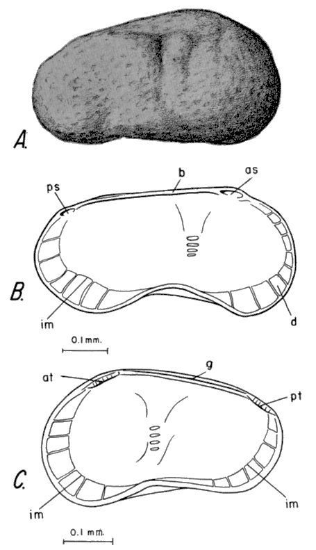
Diagnosis--The species differs from the other described species of Limnocythere by its small size, subquadrate shape, and the arched dorsal margin of the female. The lack of prominent alae is also distinctive.
Description--The female carapace is subquadrate in lateral view. The dorsal margin of the left valve is arched but the carapace exhibits a slight sinuation in the central dorsal region posterior to the middle. The arched dorsal margin of the right valve is sinuate at the junction of the valves along the central dorsal margin. The sulcus fades ventrally to the subcentral pit. The ventral margins of both valves are sinuate in the central ventral region. The anterior margins of both valves are more broadly rounded than the posterior margins. The greatest height is located anterior to the middle and is more than half the length. Nodes are common although not as pronounced as in other species. On each valve a small node is situated anterior to the sulcus in the dorsocentral region. On each valve a swelling is present posterodorsally from the sulcus. A prominent elongate longitudinal swelling is present above the sinuation in the central ventral region. The greatest breadth of the carapace is less than half the length.
The carapace of the female from the dorsal view is elliptical. The anterior end is pointed because of a flattening of the hinge margins. The sulcus is present at mid-length. The hinge flanges of both valves are projected upward to form sharp edges. The valves appear equal in size.
The duplicature is welded to the outer lamella and is widest anteriorly. Radial pore canals are small, inconspicuous, and widely spaced. The internal expression of the reticulations is clearly seen. The hingement is lophodont. The hinge of the left valve consists of an anterior tooth socket, a central dorsal curved bar, and a posterior tooth socket. The bar is formed by the knifelike extension of the hinge flange. The hinge of the right valve consists of an anterior crenulate hinge tooth, a central dorsal curved groove, and a posterior crenulate hinge tooth. The groove is formed by the hinge flange and the selvage. The muscle scars are situated on a raised platform, which is the internal expression of the external pit. The muscle-scar pattern consists of a subcentral row of four oval scars whose long axes are parallel to the length of the valves.
Sexual dimorphism is pronounced, and the male is more elongate than the female. The central dorsal margin is straight. The ventral sinuation is deeper than in the female. The central ventral elongate swelling is more pronounced than in the female. Other features are the same in both sexes.
Measurements--Adult female: holotype, left valve, length 0.52 mm, height 0.31 mm; paratype, right valve, length 0.49 mm, height 0.30 mm. Breadth of complete female paratype, 0.20 mm, length of complete paratype 0.56 mm. Adult male: allotype, left valve, length 0.63 mm, height 0.32 mm; allotype, right valve, length 0.55 mm, height 0.29 mm.
Remarks--Specimens of Limnocy there staplini are not abundant in the Pleistocene deposits of Meade County, Kansas. This species closely resembles L. sanctipatricii Brady and Robertson but does not have the prominent alae of the latter species. In Meade County, L. staplini is associated with Candona nyensis n. sp. and Cyprideis littoralis Brady. It seems to have lived in a permanent lake. The recrystallized specimens of Limnocythere found in the Pliocene deposits resemble L. staplini but they have lost many of their original features.
Repository--Specimens studied are reposited under catalogue no. 717563.
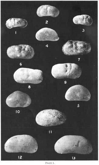
Explanation for Plate 1. All figures X60. A larger version is available as an Acrobat PDF file.
Fig. 1-3--Limnocythere staplini n. sp. 1, male left valve (allotype). 2, female left valve (holotype). 3, female right valve (paratype).
Fig. 4-5--Potamocypris smaragdina (Vávra), 1891. 4, left valve of adult. 5, right valve of adult.
Fig. 6-7--Ilyocypris gibba (Ramdohr), 1808. 6, right valve of adult. 7, left valve of adult.
Fig. 8-9--Ilyocypris bradyi Sars, 1890. 8, left valve of adult. 9, right valve of adult.
Fig. 10--Cypridopsis vidua (O. F. Müller), 1776, right valve of adult.
Fig. 11--Cypricercus tuberculatus (Sharpe), 1908, adult left valve.
Fig. 12--Candona renoensis n. sp., female left valve (holotype).
Fig. 13--Candona fluviatilis Hoff, 1942, adult left valve.
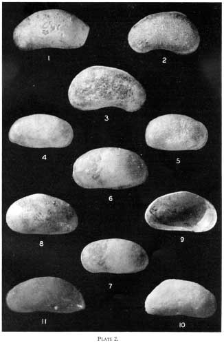
Explanation for Plate 2. All figures X50. A larger version is available as an Acrobat PDF file.
Fig. 1-3--Candona nyensis n. sp. 1, female left valve (holotype). 2, male left valve (allotype). 3, male right valve (allotype).
Fig. 4-7--Cyprideis littoralis Brady, 1889. 4, right valve of male. 5, right valve of female. 6, left valve of large male. 7, left valve of male of average size.
Fig. 8-9--Eucypris meadensis n. sp. 8, female left valve (holotype). 9, interior view of holotype.
Fig. 10--Candona crogmaniana Turner, 1894, female left valve.
Fig. 11--Candona caudata Kaufmann, 1900, female left valve.
Prev Page--Ostracode-bearing Strata || Next Page--Bibliography
Kansas Geological Survey, Geology
Placed on web November 2005; originally published June 1962.
Comments to webadmin@kgs.ku.edu
The URL for this page is http://www.kgs.ku.edu/Publications/Bulletins/157_4/04_desc.html