Kansas Geological Survey, Bulletin 38, pt. 6, originally published in 1941
Professor of Zoology, Department of Zoology, University of Kansas
Originally published in 1941 as Kansas Geological Survey Bulletin 38, pt. 6. This is, in general, the original text as published. The information has not been updated. An Acrobat PDF version (6 MB) is also available, containing parts 5 and 6.
Fossil remains of frogs and toads of the western hemisphere have been studied little, and only 7 species of extinct frogs and toads have been described from this enormous area. At least 4 frogs and toads and 1 salamander are from western Kansas. The salamanders are scarcely better known. Thus is is important to record here the presence of other salamanders, and give added information on the frogs and toads of the region, in the hope that when the entire Pliocene fauna is known, it may throw some light on the character of the climates of late Tertiary time and give some clues to the ancestry of the living amphibians of Kansas.
In this paper the following forms are described from middle Pliocene beds of extreme western Kansas: Lanebatrachus, n. gen., genotype, L. martini, n. sp. (Caudata, Ambystomidae?), Ogallalabatrachus, n. gen., genotype, O. horarium, n. sp., (Caudata, Ambystomidae), Scaphiopus antiquus, n. sp., S. pliobatrachus Taylor (Anura, Pelobatidae), Bufo hibbardi Taylor, B. arenarius Taylor (Anura, Pelobatidae. The splenial of Plioambystoma kansense Adams also is described.
One of the surprising facts concerning this fauna is the abundance of toads and the seeming absence of the ordinary frogs of the genus Rana, which today are usually the most conspicuous members of any amphibian fauna in the United States.
This is not true of the amphibian fauna of the upper Pliocene deposits of Meade County, Kansas, or the Broadwater beds of Nebraska, because in both places frogs of the genus Rana predominate, and toads are rare or absent.
The first amphibian described from the Edson beds, Ogallala formation, middle Pliocene, Sherman County, Kansas, (Adams and Martin, 1921; Elias, 1931, p. 161) was a salamander, Plioambystoma kansense Adams (Adams and Martin, 1921). This form is extremely common in these beds and several thousand separate fossil bones in nearly perfect condition have been recovered from the sand matrix. In fact, all the bony elements of the typical ambystomid salamanders have been found, except the prevomers.
Associated with this material were several fossil bones belonging to anuran amphibians. I undertook a study of these and the results were presented at the Seventh Pan-American Congress in Mexico (City), Mexico, in 1935 (Taylor, 1936). In this paper appeared the original descriptions of Scaphiopus pliobatrachus Taylor, Bufo hibbardi Taylor, and B. arenarius Taylor, which are redescribed here. Later (Taylor, 1939) I described a new pelobatid toad from a diatomaceous marl, which lies in contact with the "Edson beds" (Elias, 1931, p. 161). This was named Scaphiopus studeri Taylor.
For several years I have had at hand fragmentary fossil remains of certain other amphibians from these beds. In 1935, accompanied by David Dunkle, I visited the locality hoping to obtain additional material referable to these forms. We were unsuccessful in this; but specimens referable to two other species were obtained.
PLIOAMBYSTOMA KANSENSE Adams
In a reexamination of the material on which Adams based this species, I have noted an element that he does not mention. Eight of the lower jaws show fragments of the splenial. Inasmuch as these fragments seem to be attached to different points on the dentary, one can reconstruct the general shape and extent of the splenial bone. It is probable that these jaws belonged to neotenic or larval specimens.
The splenial is attached at a point about 1.5 mm from the symphysis on the inner side of the dentary and it extends posteriorly to a point below the last tooth of the dentary. It is somewhat curved and bears teeth, which extend the length of the element. Near the anterior point of attachment, the tooth-bearing ridge is separated from the dentary teeth by 0.6 mm. Posteriorly the splenial and dentary series are directly together. The splenial lies above the Meckelian groove and only partly covers it. This element is probably cartilaginous. In a fossilized state it is creamy white in color in sharp contrast to the darkened dentary.
The absence of prevomers and vomerine teeth in the great lot of material suggests that these elements were cartilage, and that vomerine teeth were poorly developed. It is of course not impossible that they were wholly absent from the skull.
A genus characterized by a dentary that is flattened posteriorly and bent out at an angle, and provided with a slight "coronoid" process.
This genus is named in honor of H. H. Lane, Professor of Zoology and Director of Dyche Natural History Museum, University of Kansas.
Genotype--Lanebatrachus martini, n. sp. (ambystomid salamander).
Horizon--"Edson beds", Ogallala formation, middle Pliocene, Sherman County, Kansas.
LANEBATRACHUS MARTINI, n. sp.
Figures 4A, B
Type--KUMVP no. 1468, portion of dentary and angulare, collected by H. T. Martin.
Occurrence--"Edson beds", Ogallala formation, middle Pliocene, Sherman County, Kansas.
Description--Among the material found associated with Plioambystoma kansense is a lower jaw belonging to an undescribed genus of caudate amphibians. The jaw consists of the major portion of the dentary attached to the posterior part of the angulare. There is evidence of 40 teeth along the remaining edge, 9 mm long, making a probable total number of more than 50 teeth for the complete jaw. Posteriorly the element is flattened and bent out at an angle. The greatest (vertical) width is at the point directly behind the last tooth, where there is a slight "coronoid" process. Behind this point the element narrows rapidly. The posterior tip is missing. At a point near the middle, where the jaw begins to curve inward, there is an inflation or bulge on the outer face. At no point is the jaw much thickened. Where it seems to be thicker, one notes that on the inner face the Meckelian groove is deeper. There is no trace of a splenial.
The angulare shows considerable normal torsion. The anterior higher part is turned strongly inward, and when viewed from above, it displays much more surface than this same element in Plioambystoma kansense. There is a distinct vertical groove on the thickened lower outer edge. There is no trace of the articulare.
The shape of the angulare and the dentary suggests a wide, short head, which is smaller than that of Plioambystoma kansense. The lower jaw is more delicate and more slender, and the lower edge not thickened. The dentary of Plioambystoma forms a more or less continuous curve, whereas that of Lanebatrachus has the anterior part curved inward and the posterior part bent outward, so that the main axis forms a sigmoid curve.
The species is dedicated to the late Handel T. Martin, former Assistant Curator of the Kansas University Museum of Vertebrate Paleontology, who discovered the type specimen.
A genus characterized by short vertebrae in which the centra are strongly constricted medially, and much expanded at each end.
Genotype--Ogallalabatrachus horarium, n. sp .
Occurrence--Ogallala formation, middle Pliocene, Rhinoceros Hill, Wallace County, Kansas.
OGALLALABATRACHUS HORARIUM, n. sp.
Figures 7 A-C
Type--KUMVP no. 1470, a dorsal vertebra, collected by E. H. Taylor and David Dunkle.
Occurrence--Ogallala formation, middle Pliocene, Rhinoceros Hill, Wallace County, Kansas.
Description--Associated with the vertebrae of Plioambystoma kansense, found at the above locality, is a single, large vertebra that belongs seemingly to a different genus. It probably is likewise referable to the Family Ambystomidae.
The centrum of the vertebrae is very short, but the diameter is relatively large. Posterior to the diapophyses, the centrum is greatly constricted, 1.2 mm in diameter, whereas at the anterior end it measures 4 mm, and at the posterior end 3 mm. The diapophyses and parapophyses for rib articulation arise near the middle of the bone, and are directed outward and backwards. The two are joined together almost to their distal points by a weblike bony connection, the combined width at the tip being 2.8 mm.
The length of the centrum is 5 mm. The notochordal perforation is about 0.5 mm in diameter. Within the notocoele there is a distinct offset as if a second tube were inserted in the middle of the centrum, the edges forming the offset. The prezygapophyses have large articular surfaces. The terminal neuropophysis is large and broad, bearing a small spearlike elevation that is flattened against the ventral surface and pointed backward. Two small openings occur in the tip of the neuropophysis. The elevation of the vertebra is 7 mm above the base of the centrum. The dorsal surface of the arch has a very slight keel.
Referred material--A single element, a sacral rib, is referred to this species. It is distinctly larger than the sacral rib of Plioambystoma kansense and bears a sharp spine, the tip of which is now broken. Its greatest length is 7 mm; greatest width, 3.3 mm; distance from head to base of spine, 3.2 mm.
The type specimen shows distinct torsion, and is not very symmetrical. Compared with the vertebrae of Plioambystoma the diameter of the centrum is much larger, the cup more shallow, and the centrum shorter. The notochordal canal pierces the centrum in Ogallalabatrachus horarium, which is not true of Plioambystoma kansense. I presume that this new species is a much larger form than the Plioambystoma.
The Rhinoceros Hill deposit, as interpreted by Elias (1931, p. 161) lies 50 to 80 feet higher than the "Edson beds", at the type locality of Plioambystoma. A few vertebrae, which have been temporarily referred to P. kansense, were also found in the Rhinoceros Hill deposit.
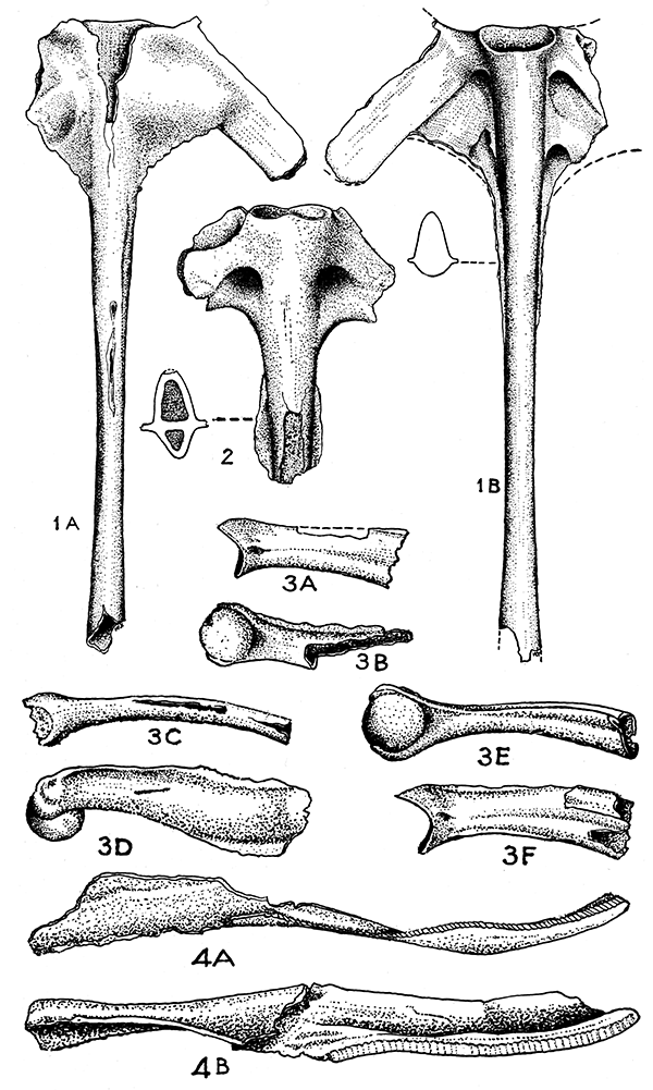
Figure 1--Scaphiopus pliobatrachus Taylor. Type (KUMVP no. 1430). Coccyx and sacral vertebra fused (actual length, 18 rom). A, Ventral view. B, Dorsal view, showing cross section.
Figure 2--Scaphiopus antiquus, n. sp. Type (KUMVP no. 1469). Sacral vertebra and coccyx fused (actual length, 7 mm).
Figure 3--Scaphiopus pliobatrachus Taylor. A, Radio-ulna (KUMVP no. 1434). B, Humerus (KUMVP no. 1432). C, Ilium (KUMVP no. 1436). D, Lateral view of humerus (KUMVP no. 1431). E, Same. F, Radio-ulna (KUMVP no. 1433).
Figure 4--Lanebatrachus martini, n. gen., n. sp. Type (KUMVP no. 1468). A, Dentary and angulare. B, Same, showing median surface (enlarged).
SCAPHIOPUS ANTIQUUS, n. sp.
Figure 2
Type--KUMVP no. 1469, portion of combined sacral vertebra and coccyx, collected by David Dunkle and E. H. Taylor.
Occurrence--"Edson beds", Ogallala formation, middle Pliocene, Sherman County, Kansas.
Description of the Type--The shaft, including bases of the sacral diapophyses, measures 5.4 mm, the total length of the fragment being 7 mm; width of the base of the sacral diapophysis, 1.3 mm; greatest diameter of the articular fossae, 1.7 mm; diameter of shaft between openings of last pair of neural foramina, 1.7 mm; greatest depth of the shaft, 2.15 mm. The seeming asymmetry of the expanded postsacral portion is due doubtless to wearing. The edges are smooth, however, as if this were the original condition. The lateral crests, now discontinuous with the expanded postsacral portion, may have been originally continuous.
Compared directly with Scaphiopus pliobatrachus there is a difference in the shape and character of the origin of the sacral diapophyses with regard to the articular fossae, and the process align="center"es of the postsacral vertebra are not in evidence. The openings for the nerves are very different and the bony web between the process and the shaft seems to be less extensive. This is shown by comparison of the measurements and drawing of the cross-section of the shaft.
The proximal width of the sacral processes in the new species described is much less than in Scaphiopus studeri Taylor, and there is distinctly more of a bony web between the transverse process and the shaft.
SCAPHIOPUS PLIOBATRACHUS Taylor
Figures 1A,B, 3A-F
Scaphiopus pliobatrachus TAYLOR, 1936, Anal. Inst. Biol. (Mexico), vol. 7, no. 4, pp. 515-521, pl. 1, fig. 1. "Edson beds", Ogallala formation, middle Pliocene, Sherman County, Kansas.
The type specimen (KUMVP no. 1430) of this species consists of a sacral vertebra joined with the coccyx. Typical of the known members of the family Pelobatidae, the coccyx is solidly fused with the sacrum. The available material shows such similarities to the modern genus Scaphiopus that I have deemed it unnecessary to erect a new genus. When complete details of the skull are known this may be necessary, however.
Description of the Type--Posterior to the widened sacral vertebrae, there seems to be evidence of a postsacral vertebra that is fused to the coccyx, as is typical in the genus Scaphiopus. No processes on the sacral element such as occur in certain living forms are seen, but the worn nature of the edges make it not certain whether they were originally absent. The coccyx is distinctly compressed and it lacks evidence of both dorsal and ventral crests. Lateral crests arise near the middle, and they widen forward, joining the expanded postsacral region, which is continuous with the proximal part of the expanded sacral diapophyses. The base of the crest is widened at the point of contact with the coccygial shaft.
The sacral vertebrae are procoelous, articulating with a single condyle. The portion of the diapophysis that is present suggests a flattened process as wide distally as its length from the centrum. The width of the diapophysis where it joins the centrum is 1.4 mm.
The neural canal is much narrowed dorsoventrally and a large antero-dorsal notch is present in the arch. The concave articular fossa is shallow, the ventral floor extending farther forward than the dorsal.
Measurements of the type in millimeters are as follows: total length of combined elements, 18; greatest depth of coccyx, at junction with sacrum, 2; width of shaft at foramina of last nerve, 2; estimated width of sacral vertebra with diapophyses, 12; width of base of the sacral diapophysis, 1.4; width of centrum between nerve foramina, 1.9.
Referred material--I am associating with this type certain other bones that seem to belong to this species-some not improbably to the same specimen, inasmuch as the proportions are such as one would postulate on the basis of measurements of modern species.
Humerus (KUMVP no. 1431). This element, which is nearly complete, shows a distinct similarity to the strongly compressed humerus of living species of Scaphiopus. The distal median condyle is globular. A strong preaxial crest (deltoid ridge) that begins near the head continues to the lateral edge of the median condyle. A well-defined postaxial crest runs nearly the length of the bone, this being absent in living forms examined. A short, low, ridge arises near the condyle below the preaxial crest. The entire element is strongly compressed. The minimum width is 1.4 mm; greatest width, 5 mm; greatest width at condyle, 3.2 mm; estimated length, 15 mm.
A second humerus (KUMVP no. 1432), a distal fragment, agrees with the previous element in many characters but is obviously from a different individual. It is somewhat smaller but the crests are precisely as in the first specimen. It has the same condylar width and its estimated length is 14.2 mm.
Radio-ulna--Two specimens of the radio-ulna (KUMVP nos. 1433, 1434) are referred to Scaphiopus pliobatrachus. The distal ends of each bone have lost the epiphyses. The postaxial (ulnar) part of each forms a moderate olecranon, and from this arises a post-axial crest that probably extends the entire length of the bones. The crest is not truly vertical from the shaft but tends to turn or curve slightly. The grooves between the ulnar and radial parts are strongly evident on both sides. Two small pitlike depressions occur near the fossa. A small lateral ridge is present on the preaxial side near the proximal end.
Specimen no. 1433 may belong to the same individual as humerus no. 1431 for they fit together perfectly. Likewise, specimen no. 1434 fits perfectly with humerus no. 1432. The estimated total length of no. 1433 is 12 mm; that of no. 1434, 11.2 mm.
Scapula--A single scapular element (KUMVP no. 1435) is referred to this species. The glenoid articular surface is narrow, elongate, curved on its inner and outer edges as is true of present day Scaphiopus. The condyle articulating with the clavicle is broken. On the dorsal edge of the head is a strong crest, rising from a point near the outer edge of the clavicular condyle. It is not vertical but slightly inclined, forming a somewhat convex surface on one side and a slightly concave surface on the other. There are no tubercles, and no subscapular depression near the dorsal proximal end is evident.
Ilium--A fragment (KUMVP no. 1436) consisting of a slightly curved shaft of an ilium broken off at the edge of the acetabulum is referred to this species. There is a trace of a groove along the inner (medial) surface. The average width is 1.3 mm. The length of the fragment is 13 mm.
Compared with Scaphiopus studeri Taylor there is a marked difference in the character of the lateral crests on the shaft of the coccyx and in the degree of distinctness of the postsacral vertebra, as well as in the union of the dilated diapophyses with the centrum. The humerus of S. studeri is longer and less widened in proportion. The two humeri measure 16 mm long by 4.6 mm wide, and 15.8 mm long by 4.8 mm wide (about 15 mm long to 5 mm wide in S. pliobatrachus); diameter of the condyle 2.3 mm (3.2 mm in S. pliobatrachus). The deltoid or postaxial crest does not extend to the distal condyle, and the other crests are not discernible.
BUFO HIBBARDI Taylor
Figure 5
Bufo hibbardi TAYLOR, 1936, Anal. Inst. Biol, (Mexico), vol. 7, no. 4, pp. 517-521, pls. 1, 2. "Edson beds", Ogallala formation, middle Pliocene, Sherman County, Kansas.
Description of Type--The specimen is complete except for the ends of the diapophyses. On the dorsal surface of the neural arch is an elevated crest forming a median right angle, from the apex of which a slight crest runs forward and downward to the anterior, interior edge of the arch. The prezygapophyses are ample, rounded, projecting, and there is a slightly hollowed area posterior to their bases. The dorsal surfaces of the diapophyses are somewhat ridged or rugose. A slight irregular ridge parallels the anterior edge of the diapophysis, behind which is a very shallow, slightly depressed area. Along the upper part of the posterior face of the diapophysis is a slight ridge, which seems to be a lateral extension of the triangular dorsal crest. The ventral surfaces of the diapophyses are smooth. The centrum bears a pair of comparatively large salient condyles, which are very closely approximated. On the ventral surface of the procoelous centrum is a longitudinal groove that becomes almost obsolete posteriorly.
The measurements of the type in millimeters are as follows: greatest transverse width (estimated), 21; actual length of longest half, 10; width of diapophyses at base, 3; estimated distal width, 8; length of centrum, 4.4; width of the two sacral condyles, 5; width of body of centrum, 4; width of arch (measured longitudinal to body; 1.9; depth of centrum, 3; height of canal, 0.9.
Referred material--Scapula (KUMVP no. 1438). This specimen is in excellent condition. The bone is slightly bowed in typical fashion. The glenoid articular surface is broad and rounded. The surface articulating with the clavicle is widened in part proximal to the fossa. On the outer face of the clavicular head of the scapula is a broad, shallow groove. Rising from the outer edge of the clavicular (acromial) head is a low, somewhat thick crest extending about a third of the length of the bone, a slight shallow groove marking its base. Below the glenoid process is a tuberclelike crest. The subscapular fossa is scarcely discernible.
Greatest length, 14.2 mm; width at neck, 3.4 mm; width at superior end, 6 mm; height from notch to end of acromial process, 4.3 mm.
Humerus (KUMVP no. 1439)--A part of the proximal end of this element, articulating with the glenoid, is missing but the distal portion is complete. The condyle is rounded, large, and the two lateral articular surfaces pronounced, each provided with a slight ridge. The distal dorsal surface of the shaft is flattened.
Width of condylar end, 6 mm; length of fragment, 17 mm; estimated total length, 32 mm.
Radio-ulna (KUMVP no. 1440). The distal articular ends of this element are missing. The fossa fits perfectly with the condyle of no. 1439. The olecranon is high and thick, the angle formed by the fossorial notch a right angle; there is a strongly defined, angular pit on the proximal anterior face. The groove between the ulnar and radial parts is evident only distally, where it is pronounced above and below. The bone is somewhat strongly compressed except at the fossorial head.
Length of fragment, 15 mm; estimated total length, 20 mm.
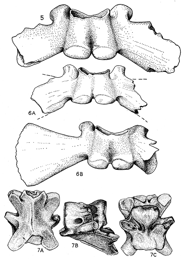
Figure 5--Bufo hibbardi Taylor. (KUMVP no. 1472). Sacral vertebra.
Figure 6--Bufo arenarius Taylor. A, (KUMVP no. 1470), sacral vertebra. B, (KUMVP no. 1474), sacral vertebra.
Figure 7--Ogallalabatrachus horarium, n. gen., n. sp. Type (KUMVP no. 1470), vertebra. A, Dorsal view. B, Same, lateral view. C, Same, ventral view. (All enlarged) .
Skull (KUMVP nos. 1441, 1442). Fragments of the posterior portion of the skull of two individuals seem to belong to the same species in spite of some variations, but they certainly are from different individuals, inasmuch as both are from the right side. The condyles and prootic regions are typical of the genus. The foramen ovale is open, the columella lacking. The knoblike projection on the medial edge of the proötic, (typical of the Bufonidae), varies slightly in character in the two specimens.
Nos. 1443, 1444, and 1445 are fragments of the frontoparietal, from which very heavy, thick crests arise. These are especially heavy at the juncture of the supraorbital and the postocular crests.
Urostyle (KUMVP no. 1446). A single large fragmentary bone is associated with this form, the dorsal anterior part, including the articular fossa, being missing. The dorsal crest rises gradually for a few millimeters, then bifurcates, forming two ridges separated narrowly along the top of the crest. How far forward this continues cannot be discerned. Anteriorly, the shaft of the urostyle becomes slightly curved in the vertical plane and flattened horizontally, forming flat shelves on each side of the crest, the edges of which project slightly. This free edge terminates forward somewhat behind a pronounced tubercle. The ventral side of the bone is finely striated.
The length of the fragment is 23 mm; the estimated total length is 27 mm.
Femur (KUMVP no. 1447). A fragment of a femur is assigned to this form. The bone is slightly sigmoid. The acetabular head is missing. A very much compressed crest arises near the middle and continues forward. At its highest elevation it presents a very sharp edge, then descends and spreads to form a flattened facet fox: muscle attachment. The epiphysis on the distal end is missing but the head is complete. In cross section it is much broader than deep. The shaft of the bone near the middle is nearly cylindrical but shows a very slightly angular elevation on one side.
The length of the fragment is 25 mm; the estimated total length is 31mm.
Tibia-fibula (KUMVP no. 1449). The shaft of this double element is nearly complete except for the epiphyses. The foramina on the shaft open on opposite sides, one about 2 mm in advance of the other.
The length of the fragment is 30 mm; the estimated total length is 33 mm.
Ilium (KUMVP no. 1449). A single complete ilium and two acetabular fragments are referred to this form (KUMVP nos. 1450,1451). The shaft is strongly curved, a partly broken, but well-defined dorsal prominence occurring above the acetabulum. A slight medianly directed crest is present on the dorsal surface near the middle of the shaft. A distinct longitudinal groove is present on the outer face. Below the acetabulum the bone is very thin and flares out somewhat. The two fragments of the acetabular portion do not differ appreciably.
The width of the basal portion is 10 mm; the length of the bone is 32 mm; the greatest diameter of the acetabular fossa is 5 mm.
Sacral vertebra (KUMVP no. 1472). A second specimen of a sacral vertebra has been obtained from the same locality as the type since the first description was published. It agrees with the type in essential details. A figure is given of this specimen (fig. 5).
The measurements in millimeters are as follows: actual length of element (termination of the diapophyses broken), 18; length of centrum, 4.5; combined width of condyles, 5.2; width of diapophyses at base, 2.9; width of body of centrum, 4.1; width of arch, measured longitudinally to body axis, 2; depth of centrum, 3.1; height of canal, 1; posterior width of canal, 3.1.
BUFO ARENARIUS Taylor
Figure 6A, B
Bufo arenarius TAYLOR, 1936, Anal. Inst. Biol. (Mexico), vol. 7, no. 4, pp. 521- 525. "Edson beds", Ogallala formation, middle Pliocene, Sherman County, Kansas.
The type of this species is a sacral vertebra (KUMVP no. 1452) collected by H. T. Martin and Claude Hibbard.
The sacral vertebra is somewhat fragmentary, lacking the greater portion of the right transverse process, and the anterior edge of the left is likewise broken. The condyles are complete and in dorsal view are only slightly visible, being concealed by the arch. They are separated from each other at their bases by a distance of about 0.4 mm. The total width across the two condyles is 3.6 mm. Each condyle is wider transversely than vertically. On the ventral surface of the centrum, anterior to the condyles, are two slightly raised, transverse areas. The groove between the condyles continues forward, but is very shallow. The vertebra is procoelous, the cavity being deep, distinctly wider transversely than vertically (2.6 rnm and 1.8 mm) .
The neural canal is much wider than deep (2 mm; 0.7 mm). The neural arch is flattened, and has a slight V-shaped crest, which is flattened on the dorsal surface. On the posterior side of the arch are two indistinct, transverse concavities or grooves. The transverse process is widened; its actual width cannot be ascertained, but, judging by extending the lines on the side of the base, it seems to be slightly more than a fourth less than the length from the point of attachment to the centrum. The posterior edge of the basal portion has a slight, irregularly roughened crest, thickened proximally and terminating in a narrow point. The prezygapophyses are represented by the broken base of the left one only.
The measurements of the type in millimeters are as follows: length of the centrum, 2.8; width of centrum anteriorly, 2.6; greatest height of vertebra, 3.1; width of constricted base of transverse process, 2.1; estimated length of transverse process to middle of centrum, 10; estimated distal width of transverse process, 5.
Referred material--Vertebra (KUMVP nos. 1453, 1454). These two vertebrae are very probably a part of the type specimen and represent the second and fourth. One is virtually complete, the other fragmentary; the processes are missing. The pre- and post-zygapophyses are of proper size for articulation together. The centra are very short (exclusive of the condyle they are slightly more than 2 mm in length); the arch is high; the cavity of the second vertebra has a vertical diameter of 2.5 mm.
Humerus (KUMVP nos. 1455, 1456). These are the distal ends of the humeri having a rounded condyle, flanked by two typical articular surfaces. The total width of the distal head is 5 mm.
Radio-ulna (KUMVP no. 1457). This element, complete except for a fragment missing from the side of the ulnar shaft, has a somewhat narrow olecranon. In lateral profile the angle of the olecranon with the head of the radium is a right angle. It fits perfectly with the condyle of no. 1455. The crest on the postaxial edge is almost completely obsolete. The groove separating the two elements is distinct. Its total length is 12 mm.
Scapula (KUMVP no. 1458). This element is complete and typical of Bufo. The end articulating with the suprascapula is moderately widened and on the ventral surface there is a somewhat broadened, shallow concavity. The acromial head is considerably longer than the glenoid process, and bears a low crest, which continues to the constricted portion of the bone. Below the glenoid process there is an elongate tubercle or crest, deflected laterally, reaching the base of the process.
Femur. Three femora (KUMVP nos. 1459, 1460, 1461) are referred to this form, the first complete except for the epiphyses. The bone is somewhat sigmoid. Posterior to the acetabular head, a low keel arises, which bears an elongate facet for muscle attachment on the anterior part. Both heads are distinctly longer than wide. No. 1460 has the constricted portion of the bone slightly narrower, and the distal head slightly larger than the preceding. The acetabular head is wanting. In no. 1461 the acetabular head and part of the epiphysis are present, as well as the anterior part of the shaft and the keel. The last two elements seem to belong to the same individual. Nos. 1462 and 1463 represent the distal halves of humeri having the epiphyses intact.
The measurments of no. 1459 in millimeters are as follows: length, 25; estimated length with epiphyses, 28; width of constricted portion, 1.4; width of shaft and crest, 3; width of distal head (no. 1463), 6.
Tibia-fibula (KUMVP no. 1464). This bone is represented by the distal half only, and has the epiphysis intact. It is much compressed, the width being 6 mm, the greatest thickness, 2.3 mm. There are two articular grooves, separated on the ventral surface by a raised tubercle. On the dorsal surface there is a narrow groove on the head. The dorsal line of the shaft is concave. The foramina on dorsal and ventral surfaces are separated by about 2 mm. The shaft is flattened, being 2 mm by 1.4 mm in its narrowest part. The groove separating the two elements is distinct on the ventral surface as far as the foramen, and to about half the distance to the tubercle, on the dorsal side.
Ilium (KUMVP nos. 1465, 1466, 1467). Two bones, complete except for the posterior tips, and belonging to the same individual, are referred. The elements are strongly curved, and have a longitudinal curved groove on the outer face. The inner face shows a curved groove near the upper border. There is a large, well-defined supra-acetabular tubercle. No. 1467 consists of the shaft only, and is from a slightly larger individual. The length of nos. 1465, 1466 is (both), 21.5 mm; the width of the head is 9 mm; the greatest diameter of the acetabular fossa is 5 mm.
Sacral vertebra. Two sacral vertebrae have been obtained from the "Edson beds" since the original description appeared. Figures of these are given (fig. 6). These agree with the type in general characters. The following measurments apply to the two sacra.
| Measurements in mm of specimens nos. 1470, 1471 | ||
|---|---|---|
| no. 1470 | no. 1471 | |
| Total width of fragment | 11.5 | 14. |
| Width of right half (complete) | ? | 10.2 |
| Length of centrum and condyle | 3. | 3.6 |
| Combined condylar width | 4.2 | 4.2 |
| Space between base of condyles | .2 | .4 |
| Width of diapophysis at base | 2.2 | 2.3 |
| Width of diapophysis at termination | ? | 7. |
| Width of centrum | 3.2 | 3.2 |
| Width of arch (measured to long axis of body) | 1.3 | 3.2 |
| Depth of centrum | 2. | 2.4 |
| Height of neural canal | 1. | 1.1 |
| Posterior width of neural canal | 3. | 3.1 |
Adams, L. A., and Martin, H. T., 1929, A new urodele from the Pliocene of Kansas: Am. Journ. Sci., 5th ser., vol. 17, pp. 504-520, figs. 24-32.
Elias, Maxim K., 1931, The geology of Wallace County, Kansas: Kansas Geol. Survey, Bull. 18, pp. 1-254, pls. 1-41. [available online]
Taylor, Edward H., 1936, Una nueva fauna de batrácios anuros del Plioceno medio de Kansas: Anal. Inst. Biol. (Mexico), vol. 7, no. 4, pp. 513-529, pls. 1-2.
Taylor, Edward H., 1939, A new anuran amphibian from the Pliocene of Kansas: Kansas Univ. Sci. Bull, vol. 25, no. 18, pp. 407-417, pls. 42-45.
This brief explanation of technical terms is given for use of readers who may not be acquainted with them.
acetabular head--Part of the thighbone that fits into acetabulum.
acetabulum--The cuplike cavity of the pelvis into which the thigh bone fits.
anchlyosed--Fused or solidly joined by bony growth.
angulare--A bone of the posterior part of the lower jaw.
Anura--A group of back boned animals that includes frogs and toads.
articulare--The posterior bone of the lower jaw.
centrum--The body of a vertebra.
coccyx--An elongate bone that terminates the vertebral column.
condyles--Slight projections from the skull with which the first vertebra is in contact.
coronoid--A small bone on the inner side of the lower jaw.
Crotaphytus--A genus of lizards, which includes the collared lizard or mountain boomer.
dentary--A bone in the anterior part of the lower jaw, which may bear teeth.
diapophyses--Bony projections from a vertebra to which a rib may attach.
diastema--A space or gap between the teeth.
epiphyses--Bony caps at the ends of the long bones, which normally fuse to the bone as the animal becomes adult.
foramina--Small openings in bones for passage of nerves and bloodvessels.
fossa--A groove or trench.
glenoid fossa--A cavity into which the bone of the upper arm fits.
matrix--The material in which a fossil is originally imbedded.
maxilla--Bone of the upper jaw.
Meckelian groove--A groove on the inner side of the dentary.
neotene--An animal that retains larval characters throughout its life.
neotenic--Having the characters of a neotene.
neuropophyses--Bony projections from the upper part of the vertebra.
notochord--A rod of cartilage that pierces the centrum of the vertebra during the early development of the larva, and mayor may not persist in the adult.
notocoele--A cuplike cavity at the anterior or posterior end, or at each end of the centrum.
olecranon process--A curved bony projection at the end of the ulna.
parapophyses--Bony projections from the sides of the vertebrae.
Phrynosoma--A genus of lizards that includes the "horned-toads".
pleurodont teeth--Teeth that are attached on the inner side of the jaw.
Pliocene--A division of geologic time. Pliocene time is estimated as having begun about 7,000,000 years ago, and ended 2,500,000 years ago.
premaxillary--A small bone in the anterior part of the lower jaw.
prevomer--A bone in the roof of the mouth.
prezygapophyses--Bony processes from the arch of the vertebra.
procoelus--Referring to a vertebra having a cuplike notocoele at the anterior end.
pulmonate mollusca--A group of invertebrate animals that include snails.
radio-ulna--The bone of the arm, formed by the fusion of the radius and ulna.
sacral vertebra--The vertebra to which the ilia attach.
sigmoid--Having an S-shape.
splenial--A bone of the inner side of the lower jaw.
symphysis--The line of union between two bones.
urostyle--A bone terminating the vertebral column; the coccyx.
Kansas Geological Survey
Placed on web March 18, 2015; originally published July 7, 1941.
Comments to webadmin@kgs.ku.edu
The URL for this page is http://www.kgs.ku.edu/Publications/Bulletins/38_6/index.html