Kansas Geological Survey, Bulletin 38, pt. 5, originally published in 1941
Professor of Zoology, Department of Zoology, University of Kansas
Originally published in 1941 as Kansas Geological Survey Bulletin 38, pt. 5. This is, in general, the original text as published. The information has not been updated. An Acrobat PDF version (6 MB) is also available, containing parts 5 and 6.
The state of Kansas is widely known for its fossil-bearing rock strata, which have yielded a great number and variety of extinct vertebrate animals that lived in past ages. For the most part, the vertebrate fossils that are best known belong to the larger forms of mammals, reptiles, and fishes, inasmuch as these are usually the first to be collected. This is probably due to the fact that fossilized bones of the larger animals are more readily discovered and more easily excavated. The faunas of the past, however, did not consist merely of large animals. Just as today we find small species living in the same regions with the larger species, so we may expect the same to be true of past geological periods or epochs.
I have undertaken to find out something about the varieties of these small forms, particularly, the species of lizards, snakes, frogs, toads, and salamanders that lived during late Pliocene time in Kansas; to ascertain their relationships with the present living forms; and to determine whether they throw any light on the climatic conditions that existed in the past, when camels, rhinoceroses, peccaries, three-toed horses, and mastodons were typical large mammals of the state.
This paper treats only of the lizards of the upper Pliocene deposits in Kansas. Four species are here reported for the first time. That this is a significant number is evident when we realize that only three species of lizards have been heretofore known from all of Miocene, Pliocene, and Pleistocene time in North America. One of the three is a horned-toad, one a collared lizard, and these two have been referred to the living genera Phrynosoma and Crotaphytus, respectively; one, which is from the Miocene, has been made the type of a new genus, having no known living representatives.
In this paper one genus and four species of lizards are described from the upper Pliocene beds of Meade County, Kansas. These are Eumecoides, n. gen. (Scincidae?), genotype E. hibbardi, n. sp., E. mylocoelus, n. sp., Eumeces striatulatus, n. sp. (Scincidae), and Cnemidophorus bilobatus, n. sp. (Teiidae).
The first two species are small lizards whose relationship is not clear. They are placed in a new genus, and referred, with some doubt, to the family of the skinks. The third species is a skink of the widely distributed genus Eumeces, which has living representatives in Africa, Asia, and North America. The fourth species is a whiptail lizard, belonging to a living genus, and to a family which is confined to the western hemisphere.
A small collection of reptilian fossils from the upper Pliocene beds of Meade County, Kansas, was placed in my hands for study by Claude Hibbard, Assistant Curator of Paleontology, in the University of Kansas Museum. The specimens were collected by Hibbard and his party during the summers of 1938 and 1939, and are now a part of the collections of the Kansas University Museum of Vertebrate Paleontology (KUMVP).
The material consists, for the most part, of jaw elements. In no case were any of the separate remains closely associated, so that there is no assurance that any individual is represented by more than a single element. Hibbard regards the beds that yield these fossils as being a flood-plain deposit. The matrix consists chiefly of sandy silts and clays.
Associated with the lizards are jaws and vertebrae of snakes, at least one salamander, several birds, and numerous mammals. A few invertebrate animals, chiefly pulmonate mollusca, are present in the deposit. The contemporary mammalian fauna is being studied by Hibbard, who finds that few of the living genera of mammals are represented.
C. M. Gilmore (1928) has listed a single genus and species of lizard (Phrynosoma?) as occurring in Pleistocene deposits of North America, and a single genus and species (Crotaphytus?) in the Pliocene. Nothing is listed for the Miocene, but a new saurian genus, Tetralophosaurus, has recently been proposed by Olson (1937) based on remains from the lower Harrison beds, of Miocene age, in Nebraska.
The forms identified in recognized living genera offer little data for determining past climatic conditions, inasmuch as both genera are widespread, occupying arid and semiarid, as well as moist and wet regions. Both genera are represented in tropical and temperate regions. Both genera have representatives extending from sea level to 6,000 feet (Cnemidophorus) and 12,000 feet (Eumeces).
CNEMIDOPHORUS BILOBATUS, n. sp.
Figures 1A-C, 2A-C
Type--KUMVP no. 5084, portion of dentary containing 8 teeth, collected by Claude W. Hibbard and party, 1938.
Occurrence--Rexroad formation (Smith, 1941, pp. 95-99) upper Pliocene, Loc. 2, about 16 miles southwest of Meade, Meade County, Kansas.
Description of Type--The outer face of the type specimen shows no part of the coronoid but the scar of attachment is indicated by a slightly depressed area, extending forward to the middle of the fourth tooth (from last). Below the diastema between the eighth and ninth teeth (from last) is a forward-directed foramen, which is situated above the level of the middle of the dentary. The region toward the ventral surface, lying below the fifth to eighth teeth, is smooth, lacking any distinct striation or rugosity. On the inner face of the dentary the Meckelian groove, which is lateral (but becoming ventral anteriorly), and the Meckelian cavity are completely exposed, owing to the absence of the thin splenial, which covers the groove.
The teeth are pleurodont and unequally bilobed, the small lobe being anterior. The second, fourth, and fifth teeth (from last) show a very faint trace of a still smaller posterior notch or lobe, which is a counterpart of the third lobe, present in certain living species of Cnemidophorus. The teeth are wider in proportion to length when compared with modern representatives of Cnemidophorus living in this southwestern region, and the crowns seem more blunt, but the latter difference may be due to age and wear. The relative size of the teeth and their characters are shown in figure 1.
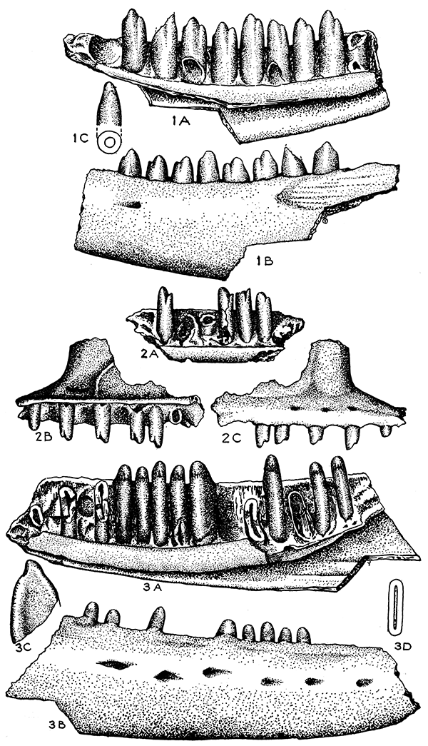
Figure 1--Cnemidophorus bilobatus, n. sp. Type (KUMVP no. 5084). A, Median view of the left dentary, enlarged. B, Lateral view of the left dentary. C, Single tooth from near the middle of the series and diagrammatic cross section.
Figure 2--Cnemidophorus bilobatus, n. sp. Referred material. A, Fragment from near the anterior tip of the left dentary (KUMVP no. 5091), enlarged. B, Fragment of the anterior part of right maxilla (KUMVP no. 5081), median view, enlarged. C, Same, lateral view.
Figure 3--Eumeces striatulatus, n. sp. Type (KUMVP no. 5079). A, Median view of the right dentary, enlarged. B, Same, lateral view. C, Single tooth from near middle of series. D, Cross section of the same.
The jaw of this extinct species has been compared with the jaws of the living Cnemidophorus sexlineatus (Linné), C. perplexus Baird and Girard, C. gularis Baird and Girard, and C. melanostethus Cope. Actually, the only one of these species known in this territory today is C. sexlineatus. This form agrees in the normal absence of distinctly trilobed teeth, but differs in having more slender and somewhat more tapering, laterally compressed teeth. The bases are proportionally closer together and the upper parts are somewhat more widely separated. In C. gularis, living in Texas, the four or five posterior teeth are generally trilobed, the posterior lobe being nearly as distinct as the anterior. The posterior teeth of C. melanostethus are more like those of C. sexlineatus, and the outer posterior face of the dentary is very definitely sculptured and somewhat rugose. C. perplexus has teeth that are heavier on the basal portions, and the posterior part of the dentary is smooth. Also, the anterior extension of the coronoid is shorter, reaching forward only to the level of the third tooth, and it is more pointed. The teeth taper more towards their tips and their bases are closer together than in the described new species.
Measurements of the type specimen in millimeters, are as follows: extreme length of fragment figured, 7; elevation of dentary at posterior lateral foramen, 1.8.
Remarks--The estimated total length of the lower jaw of this species is 25 mm, based on proportional measurements in living species. This jaw size suggests a species somewhat larger than any of the four mentioned forms.
Referred material--A fragment of a right maxilla (KUMVP no. 5081) from the type locality is referred to this species. The teeth are very considerably smaller than those of the type specimen, which are posterior dentary teeth. However, in nine living species examined, the anterior maxillary and dentary teeth are much smaller than the posterior teeth in the same jaw. In many specimens the tooth at or near the maxillary-premaxillary suture is smaller than the adjacent teeth.
The anterior unbroken profile of the fragment is the posterior outline of the nasal opening. The smallest anterior tooth seemingly is the one that follows the tooth at the suture between the premaxillary and maxillary. The outer face shows three foramina. On the medial face the maxillary shelf is extended above the base of the teeth, and a broken curving edge of a maxillary process is evident.
The teeth are bilobed, hollow, slightly compressed at the tips, but they differ from the living species in that they are almost straight rather than curved medially. The total length of the maxillary fragment is 4 mm, which includes space for 10 teeth; whereas in a space of 4 mm on the posterior dentary series, there are 7 teeth. C. gularis has 13 and 7% teeth in 4 mm in these same regions. C. sexlineatus has 12 and 8% teeth correspondingly in this distance. The measurements given are based on study of one skull belonging to each species. It is seen that the smaller maxillary teeth might be from an individual as large as the type.
EUMECES STRIATULATUS, n. sp.
Figures 3A-D, 4A,B
Type--KUMVP no. 5079, major portion of right dentary, bearing eight complete teeth and broken parts of seven more or spaces for them.
Occurrence--Rexroad formation, upper Pliocene, Loc. 2, about 16 miles southwest of Meade, Meade County, Kansas.
Description of Type--The outer face of the dentary is smooth except for depressions formed by a linear series of six somewhat large foramina, the most posterior one of which is opposite the fifteenth tooth. The outer face of the dentary is strongly curved. The bone is relatively thick, and the lower edge curves slightly.
On the median side, the large Meckelian cavity and groove are evident, the latter being almost wholly ventral. The groove, which is deep posteriorly, becomes shallow anteriorly and terminates near the symphysis. The upper medial edge of the dentary tends to fold up along the base of the teeth, making a slight trough. The lower, outer edge of the dentary is not upturned to present a medial face. The splenial is missing.
The teeth are strongly compressed transversely, each being closely anchylosed to the outer (lateral) dentary wall, so that the connections reach even above the general alveolar edge. The outer face of the tip of the tooth is somewhat rounded and curved medially. The inner, medial face of the tip is slightly concave and striated or puckered, the transverse, cutting edge being blunt.
There are 8 complete teeth in the dentary, the bases of 5 broken teeth, and spaces for 4 more. The posterior part of the dentary, bearing 4, or possibly 5 teeth, is missing. The total number of teeth is about 21, which is about the same as in many living forms. There are 6 or 7 teeth in a space of 4 mm.
Measurements of the type, in millimeters, are as follows; total length of fragment, 9; elevation of dentary at 14th tooth, 2.8; height of same tooth, 2.
Comparisons--At the present time Eumeces obsoletus Baird and Girard, E. fasciatus (Linne), E. septentrionalis (Baird), and E. multivirgatus (Hallowell) occur in territory not far distant from the type locality of the species herein described. The described species is distinctly larger than E. fasciatus, E. septentrionalis and E. multivirgatus and there is no need to compare it with these. A very large specimen of E. obsoletus may equal the size of E. striatulatus.
Compared with a moderately large specimen of E. obsoletus the following differences are seen in E. striatulatus. The teeth are proportionally much wider and somewhat longer. In a space of 4 mm there are 6 or 7 teeth as compared with 9 to 10 teeth in a similar space in E. obsoletus. The general character of the teeth is very similar, however. The shape and character of the dentary is similar also, except that the foramina are more numerous (3 only in E. obsoletus) and the anterior tip is proportionally higher.
Referred material--A few fragments are referred to this species. These include no. 5080, which is the tip of a dentary showing parts of 10 teeth or spaces for them. The tip is slightly lower than in the type. No. 5129C is the posterior fragment of a maxilla bearing 2 teeth. No. 5079A is an anterior maxillary fragment bearing several perfect teeth. These are slightly heavier than those in the type dentary and are somewhat longer. This element is, I believe, certainly from an individual other than the type.
Description--The medial ventral edge of the dentary is turned up, causing the Meckelian groove to be medial rather than ventral. The anterior end of the dentary is narrowed. There are 19 to 22 closely set pleurodont teeth that are strongly anchylosed to the outer wall. The teeth are hollow, having a blunt, chisel-like tip. They are compressed laterally, widening more or less toward the base. Striations are lacking on the medial face of the tips. Referred to Scincidae (with some doubt).
Genotype, Eumecoides hibbardi, n. sp.
Two forms, each represented by dentaries, are referred to the genus. They may be differentiated by the following key.
A. Outer face of dentary concave; terminal part of Meckelian groove shallow; about 18 mandibular teeth. Teeth moderately compressed laterally …
E. hibbardi, n. sp.
B. Outer face of dentary convex; terminal part of Meckelian groove deeper; 21 to 23 mandibular teeth; teeth strongly compressed laterally …
E. mylocoelus, n. sp.
EUMECOIDES HIBBARDI, n. sp.
Figures 5A-D
Type--KUMVP no. 5099, a virtually complete dentary, containing 15 teeth and spaces for 3 more. Collected by Claude W. Hibbard and party, 1936.
Occurrence--Rexroad formation, upper Pliocene, Loc. 2, about 16 miles southwest of Meade, Meade County, Kansas.
Description of Type--The dentary tapers gradually toward the anterior end, where the width is slightly less than a third of the posterior width. A very slight constriction occurs just back of the first (anterior) tooth. The outer face of the dentary shows a shallow, but distinct, longitudinal concavity. There are 7 foramina, which are not arranged regularly but in a more or less linear series on the anterior part, the posterior foramen being opposite the base of the 10th tooth. The posteroventral surface of the dentary is more than one-half of the width of the outer face, narrowing anteriorly. The medial edge of the ventral part of the dentary is turned up, causing the Meckelian groove to open medially rather than ventrally. The greatest width o{ the upturned edge is anterior to the middle. The groove is almost obsolete at the top. The medial face of the dentary is turned up, so as to conceal the bases of the teeth, in lateral view, its widest part being at about the middle. No trace of a scar or suture of the coronoid is seen, and the splenial suture hardly is evident.
The teeth are of the pleurodont type, those near the median part of the series being longest and most robust. There are about 18 hollow teeth (3 or possibly 4 missing) , their tips being somewhat compressed longitudinally, so as to be bluntly chisel-like, and each tooth as a whole is more or less compressed transversely to the long axis of body (fig. 5C). Anchylosis of the tooth and bone takes place below the alveolar border, instead of reaching to and above the alveolar level as in Eumeces striatulatus. The narrowest diameter of the teeth is much less than one-half of the greatest diameter.
The species is named for Claude W. Hibbard, Curator of Lower Vertebrates, Dyche Museum of Natural History, University of Kansas.
Measurements of the type specimen, in millimeters, are as follows: greatest length of dentary, virtually complete, 8.2; greatest vertical width, 2.65; narrowest point, posterior to first tooth, .75, greatest ventral width, 1.5; height of longest tooth, 1.15.
Remarks--The reference of this genus to the Scincidae is based on general resemblances more than on specific characters. The relationship to Eumecoides mylocoelus, n. sp., is shown in the key.
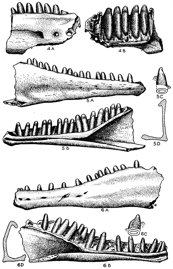
Figure 4--Eumeces striatulatus, n. sp. Referred material. A, Fragment of right dentary from near middle of bone, lateral view, enlarged. B, Same, median view.
Figure 5--Eumecoides hibbardi, n. gen., n. sp. Type (KUMVP no. 5099). A, Lateral view of right dentary, enlarged. B, Same, medial view. C, Tooth from middle of series. D, Cross section of posterior part of dentary.
Figure 6--Eumecoides mylocoelus, n. gen., n. sp. Type (KUMVP no 5115). A, Lateral view of left dentary, enlarged. B, Same, medial view. C, Single tooth from near middle of series. D, Cross section of dentary (reconstructed to show contrast with fig. 5D).
EUMECOIDES MYLOCOELUS, n. sp.
Figures 6A-D
Type--KUMVP no. 5115, a dentary slightly broken posteriorly, bearing spaces for about 23 teeth (15 teeth present). Collected by Claude W. Hibbard and party, 1936.
Occurrence--Rexroad formation, upper Pliocene, Loc. 2, about 16 miles southwest of Meade, Meade County, Kansas.
Description of Type--The outer face of the dentary is generally convex, but two slight, longitudinal depressions are evident on the posterior part. A longitudinal series of 5 somewhat large foramina occupies the anterior part of the outer face, nearly midway between the upper and lower edges. The most posterior of the series is about opposite the base of the 11th tooth. The lower profile of the dentary is somewhat sinuous, a prominent elevation on the anterior ventral border being opposite the third tooth. The element is widest posteriorly, narrowing to a point between the fifth and seventh teeth, where the greatest constriction occurs. The greatest width of the dentary is about 2.5 times the narrowest width. The greatest width of the ventral surface is more than one-half of the greatest width of the lateral surface. No trace of the coronoid suture is seen. The medial ventral edge, distinctly thickened, is turned up on the medial side, causing the Meckelian groove to present a median rather than a ventral opening. The groove continues to be well defined to the anterior tip of the dentary. The median face of the dentary above the groove is hardly upturned, at least on the anterior half (broken posteriorly), so that in lateral view the tooth bases are not concealed by an upturned edge.
Tips of the teeth are compressed, forming a blunt chisel-like edge longitudinally, but the teeth are strongly compressed transversely to the longitudinal axis, the greatest diameter of each tooth being twice its narrowest diameter. The teeth have a narrow, oval pulp cavity. Teeth of the medial part of series are longer and most are wider transversely than the other teeth. The anchylosis of the tooth and bone does not extend quite to the upper level of the dentary bone.
Measurements of the type, in millimeters, are as follows: total length of dentary, 8.6 (possibly slightly longer if complete); greatest vertical width, 2.45 (possibly 4.6 if complete); narrowest width between fifth and seventh teeth, .95; greatest ventral width, 1.3; height of longest tooth, 1.2.
Remarks--The chief difference between the teeth of the two forms referred to this genus is the greater width of the teeth in Eumecoides mylocoelus. The teeth illustrated are from the same place in the jaws, that of E. mylocoelus being the twelfth, and that of E. hibbardi, the tenth tooth. However, I do not deem these differences greater than differences that obtain in species of other living genera of saurians.
Gilmore, Charles W., 1928, Fossil lizards of North America: Nat. Acad. Sci., Mem., vol. 22 (Mem. 3), pp.1-169, pls. 1-27.
Olson, Everett Claire, 1937, A Miocene lizard from Nebraska: Herpetologica, vol. 1, no. 4, pp. 11-112, fig. 1.
Smith, H. T. U., 1941, Geologic studies in southwestern Kansas: Kansas Geol. Survey Bull. 34, pp. 1-244, pls, 1-33, figs. 1-22.
This brief explanation of technical terms is given for use of readers who may not be acquainted with them.
acetabular head--Part of the thighbone that fits into acetabulum.
acetabulum--The cuplike cavity of the pelvis into which the thigh bone fits.
anchlyosed--Fused or solidly joined by bony growth.
angulare--A bone of the posterior part of the lower jaw.
Anura--A group of back boned animals that includes frogs and toads.
articulare--The posterior bone of the lower jaw.
centrum--The body of a vertebra.
coccyx--An elongate bone that terminates the vertebral column.
condyles--Slight projections from the skull with which the first vertebra is in contact.
coronoid--A small bone on the inner side of the lower jaw.
Crotaphytus--A genus of lizards, which includes the collared lizard or mountain boomer.
dentary--A bone in the anterior part of the lower jaw, which may bear teeth.
diapophyses--Bony projections from a vertebra to which a rib may attach.
diastema--A space or gap between the teeth.
epiphyses--Bony caps at the ends of the long bones, which normally fuse to the bone as the animal becomes adult.
foramina--Small openings in bones for passage of nerves and bloodvessels.
fossa--A groove or trench.
glenoid fossa--A cavity into which the bone of the upper arm fits.
matrix--The material in which a fossil is originally imbedded.
maxilla--Bone of the upper jaw.
Meckelian groove--A groove on the inner side of the dentary.
neotene--An animal that retains larval characters throughout its life.
neotenic--Having the characters of a neotene.
neuropophyses--Bony projections from the upper part of the vertebra.
notochord--A rod of cartilage that pierces the centrum of the vertebra during the early development of the larva, and mayor may not persist in the adult.
notocoele--A cuplike cavity at the anterior or posterior end, or at each end of the centrum.
olecranon process--A curved bony projection at the end of the ulna.
parapophyses--Bony projections from the sides of the vertebrae.
Phrynosoma--A genus of lizards that includes the "horned-toads".
pleurodont teeth--Teeth that are attached on the inner side of the jaw.
Pliocene--A division of geologic time. Pliocene time is estimated as having begun about 7,000,000 years ago, and ended 2,500,000 years ago.
premaxillary--A small bone in the anterior part of the lower jaw.
prevomer--A bone in the roof of the mouth.
prezygapophyses--Bony processes from the arch of the vertebra.
procoelus--Referring to a vertebra having a cuplike notocoele at the anterior end.
pulmonate mollusca--A group of invertebrate animals that include snails.
radio-ulna--The bone of the arm, formed by the fusion of the radius and ulna.
sacral vertebra--The vertebra to which the ilia attach.
sigmoid--Having an S-shape.
splenial--A bone of the inner side of the lower jaw.
symphysis--The line of union between two bones.
urostyle--A bone terminating the vertebral column; the coccyx.
Kansas Geological Survey
Placed on web March 18, 2015; originally published July 7, 1941.
Comments to webadmin@kgs.ku.edu
The URL for this page is http://www.kgs.ku.edu/Publications/Bulletins/38_5/index.html