


Ancient Life in Kansas Rocks, part 21 of 27
Crinoids, various fragments of which are pictured here slightly enlarged, are known from rock formation from the Lower Ordovician to the Recent. They have living representatives with world-wide distribution. Exclusively marine, these highly complex and varied echinoderms are commonly found as dissociated, broken apart and scattered, fragments. Often referred to as "sea lilies,", most crinoids resembled flowers and sometimes grew in garden-like clusters.
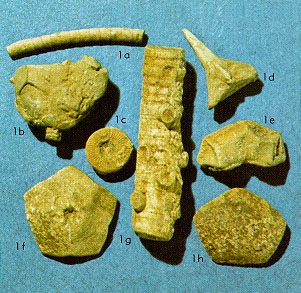
Most commonly found are fragments of the stem, represented as long slender fragments (1a and 1g), or isolated columnal plates, the round button-like fragment (1c). The stem or column is composed of many columnals stacked one above the other. Columnals commonly have radiating ridges called crenella, and a central hole, the lumen (1c). The lumens together comprise an axial canal down the center of the stem that contains some of the animals organs. The columnals may interlock or may have ligamentous tissue inserted between the facets allowing varying degrees of flexibility (some what like the backbone of a human).
Columnals may be circular to pentagonal, star-shaped or elliptical, and may be wider than long to longer than wide. Many stems thicken and branch at the base (resembling a root) and hold the animal fast to the soft substrate; however, the animal does not receive nutrition through the root as do plants, but rather has a complex food-gathering system in the crown. The crown is situated at the top of the stem and is composed of the dorsal cup, arms, and tegmen (see drawing below). 1b in the photograph is a dorsal cup with a portion of its stem still affixed. Note that it is composed of plates that fit closely together and form a cup. It is within this cup that the vital organs are enclosed. 1e, 1f, and 1h are plates from such a cup.
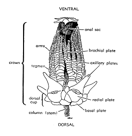
Reaching above the cup are arms composed also of minute plates variously arranged in elongate series. The plates in the cup to which the arms attach are termed ray plates or radials. The plates in the arm series are termed brachial plates. The arm continues upwards with a series of brachials until it divides at a specialized brachial plate, an axillary (see drawing). The spinose brachial plate (1d in the photograph) is an axillary plate, as shown in the drawing. Above the axillaries continue a series of brachials, then dividing at axillaries and so on.
Different crinoids may have all or only a few of these features. Commonly set between two radials is a special plate that leads to the anal series forming an anal sac or tube. In the drawing above, the arms are not shown on the left side to reveal the sac that rises above the tegmen. The tegmen is an area of the crown above the cup and within the arms, covered by a leathery membrane or by plates. Down the inner surface of the arms and across the tegmen to the mouth are depressions termed ambulacral grooves. Along these grooves are located cilia that generate water currents to carry food along the grooves to the mouth. (Lecompton Limestone, Upper Pennsylvanian)
Kansas Geological Survey
Placed online Feb. 1997
URL = "http://www.kgs.ku.edu/Publications/ancient/f21_crin.html"
Send comments and/or suggestions to webadmin@kgs.ku.edu